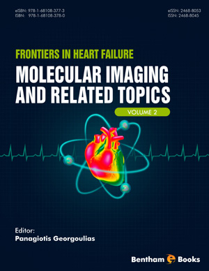
Book Volume 2
List of Contributors
Page: vii-ix (3)
Author: Panagiotis A. Georgoulias
DOI: 10.2174/9781681083773116020004
Computed Tomography in Heart Failure
Page: 3-25 (23)
Author: Ioannis A. Chryssogonidis, Iokasti E. Gkogkou and Christos A. Papadopoulos
DOI: 10.2174/9781681083773116020005
PDF Price: $30
Abstract
Multidetector computed tomography is an imaging modality which constantly gains ground in the field of cardiovascular imaging. Accurate imaging of coronary arteries, cardiac structure and function, pulmonary and cardiac venous anatomy can be explored with this non-invasive, easily reproducible technique, providing valuable information in the diagnosis and management of patients with heart failure. Computed tomography can support invasive techniques used in patients with heart failure, as in cardiac resynchronization and ablation for atrial fibrillation, with all necessary anatomic data, increasing the safety and efficacy of the procedures. In addition, patients after heart transplantation can be evaluated with multidetector computed tomography, avoiding more invasive procedures.
Magnetic Resonance Imaging in Heart Failure
Page: 26-67 (42)
Author: Maria A. Mademli and Nikolaos L. Kelekis
DOI: 10.2174/9781681083773116020006
PDF Price: $30
Abstract
Heart failure (HF) may be the endpoint of various cardiac diseases. This emphasizes the need for a diagnostic method that can accurately establish the diagnosis of HF and define the underlying mechanisms of the condition. This is necessary for the selection of the appropriate therapeutic method (medication, revascularization or resynchronization therapy) in order to achieve the optimal response. Cardiac magnetic resonance imaging (MRI) is an emerging imaging method that can be used for quantification of ventricular function, as a baseline and also for follow-up of HF patients after treatment. It has great contribution in differentiation of the cardiac diseases (e.g. ischemic and non-ischemic cardiomyopathies, cardiomyopathies with myocardial hypertrophy, etc.) by providing tissue characterization. This chapter describes the technique of cardiac MRI examination and focuses on typical imaging characteristics that aid in the differentiation among cardiac conditions that may result in HF, according to their frequency and their importance in treatment selection. The parameters measured by cardiac MRI that play a role in the prognosis of the disease and therapeutic decision, are also discussed. Finally we present the role of cardiac MRI in cardiac resynchronization therapy and its predictive role in the therapeutic outcome.
Myocardial Perfusion (SPECT) Imaging: Radiotracers and Techniques
Page: 68-123 (56)
Author: Panagiotis A. Georgoulias, George C. Angelidis, Athanasios S. Zisimopoulos and Ioannis C. Tsougos
DOI: 10.2174/9781681083773116020007
Abstract
Heart failure remains a highly prevalent disease with significant morbidity and mortality. Millions of patients are affected worldwide and their treatment is associated with a significant cost for the healthcare systems, especially in developed countries. In approximately two-third of the cases, ischemic heart disease is the cause of the syndrome. Therefore, myocardial perfusion single photon emission computed tomography (SPECT) imaging is a key element in the diagnostic investigation, prognostication, and management of patients with heart failure. Perfusion images are obtained according to various well-established protocols, after the administration of either thallium-201 or technetium-99m labelled radiotracers in combination with several stress techniques. In the future, technological innovations and improvements of reconstruction methods are expected to strengthen the role of myocardial perfusion SPECT imaging as a useful tool for the investigation of heart failure and patient’s management.
Radionuclide Assessment of Cardiac Function and Μodeling: The Clinical Application of Gated- SPECT
Page: 124-151 (28)
Author: Spyridon Tsiouris, Athanasios Papadopoulos and Andreas Fotopoulos
DOI: 10.2174/9781681083773116020008
PDF Price: $30
Abstract
Nuclear myocardial perfusion imaging (MPI) by single-photon emission computed tomography (SPECT) is a non-invasive method of evaluating left ventricular (LV) perfusion and viability that is widely used in clinical practice. Electrocardiographically (ECG)-gated SPECT (G-SPECT) is a state-of-the-art technique combining the evaluation of myocardial perfusion and LV function within a single study. It is among the most commonly performed cardiologic diagnostic procedures in nuclear medicine departments. Advances in γ-camera instrumentation and the software used for data acquisition, image processing and quantification have rendered this technique user-friendly, practical and highly reproducible in everyday practice. In patients with coronary artery disease, the introduction of ECG-gating enhances the diagnostic and prognostic capability of MPI, provides incremental functional information over the perfusion data alone and also bears potential for assessing myocardial viability and following-up LV function after revascularization. This chapter discusses the general principles of G-SPECT image acquisition, analysis and quantification, followed by a discussion on the additive diagnostic and prognostic value that is associated with the functional parameters obtained by G-SPECT.
Evaluation of Heart Failure Patients Using PET Perfusion Imaging: Radiotracers and Techniques
Page: 152-171 (20)
Author: Sofia Chatziioannou, Nikoletta Pianou and Alexandros Georgakopoulos
DOI: 10.2174/9781681083773116020009
PDF Price: $30
Abstract
Positron emission tomography has become an increasingly alternative method for clinical application in patients with coronary artery disease. Although, various myocardial perfusion PET tracers are available, the most commonly used in clinical setting are Rubidium-82 (82Rb) and nitrogen-13-ammonia (13Ν-ammonia). 82Rb is a cation and an analog of potassium with kinetic properties similar to those of Thallium-201. It combines the advantages of a short 75 seconds physical half-life and its independency of an onsite cyclotron through the availability of a relative small onsite Strontium-82 / 82Rb generator. Although it is not the ideal tracer for absolute quantification (first–pass extraction of 65%), it has been used extensively for this purpose showing valuable data in the clinical setting. 13Ν-ammonia is a cyclotron product with physical half-life of 9.96 minutes. Due to the combination of the high first-pass myocardial extraction fraction (80%) and the relatively long physical half-life of the radiotracer, high-contrast resolution myocardial perfusion images can be obtained. Oxygen -15-water (15O-water) is used in research studies, mainly for precise measuring of myocardial blood flow. 18F-Fluripiridaz is a new promising tracer with excellent biological and imaging characteristics, including longer half-life, availability in unit doses from regional cyclotrons, low positron range, and high myocardial extraction. It is now in advanced clinical evaluation with encouraging results. Due to the short half-life of the radiotracers a PET rest-stress study is obtained in a shorter time than a single photon emission computed tomography study, while PET myocardial perfusion imaging provides higher diagnostic accuracy, using lower radiation doses compared to single photon emission tomography.
Hybrid Imaging (SPECT/ CT, PET/CT, PET/MR)
Page: 172-239 (68)
Author: Dimitrios J. Apostolopoulos
DOI: 10.2174/9781681083773116020010
PDF Price: $30
Abstract
Multi-modality imaging achieves the integration of structural and functional or metabolical information in a single examination. Apart from patient convenience and improved workflow, this “one-stop shop” approach is featured by enhanced diagnostic accuracy compared to either modality alone or side-by-side image interpretation. These advantages also apply on cardiovascular and molecular-targeted imaging where hybrid systems facilitate the detection of molecular signals and their accurate localization by fusion with anatomical structures. The role of SPECT/CT, PET/CT and PET/MR in studying patients with heart failure is reviewed in this chapter. Before mentioning the potential clinical utility, various issues concerning the principles of hybrid imaging, commercially available devices, image interpretation, possible technical errors and diagnostic pitfalls are addressed. Due to its wider availability, lower cost and the author’s experience, the value of cardiac hybrid SPECT/CT is emphasized.
Assessment of Myocardial Viability Using SPECT and PET Techniques
Page: 240-299 (60)
Author: Efstratios Moralidis
DOI: 10.2174/9781681083773116020011
PDF Price: $30
Abstract
Heart failure is a significant health problem and coronary artery disease is by far the leading cause. Despite advances in medical and device therapy the prognosis of patients with ischemic cardiomyopathy remains unfavorable, but revascularization may further improve the outcome in terms of contractile function, symptomatic relief, exercise capacity and mortality. Over the years, the presence of myocardial viability has been considered a significant determinant of the benefit from revascularization and a variety of noninvasive techniques have been developed to assess viable and nonviable myocardium in patients with ischemic systolic dysfunction. Viability imaging with 201Tl and 99mTc-agents SPECT can evaluate perfusion, cell membrane and mitochondria structural and functional integrity, whereas 18F-FDG PET is used for the assessment of glucose metabolism in myocytes. Dobutamine stress echocardiography provides information on the contractile reserve and cardiac magnetic resonance imaging can delineate the transmural extent of scar. In general nuclear imaging techniques have a higher sensitivity for the detection of myocardial viability, whereas techniques evaluating contractile reserve display a lower sensitivity but a higher specificity. This review focuses primarily on the radionuclide modalities for the assessment of myocardial viability and discusses the clinical value of viability imaging, including earlier retrospective work and the more recent prospective data.
Radioisotopic vs Non-Radioisotopic Methods for Myocardial Viability Identification
Page: 300-334 (35)
Author: Varvara I. Valotassiou and Julia V. Malamitsi
DOI: 10.2174/9781681083773116020012
PDF Price: $30
Abstract
Left ventricular (LV) systolic dysfunction associated with coronary artery disease (CAD) comprises a major diagnostic and therapeutic dilemma. Hibernating myocardium refers to a chronic dysfunctional condition, as a result of repeated episodes of ischemia, of a still viable myocardium. In viable dysfunctional myocardium, the integrity of myocyte membrane and contractile fibers are preserved. Revascularization may promote LV function in cases of residual myocardial viability in dysfunctional segments of the heart. The identification of viability is pivotal for patients’ management, and viability testing is a valuable tool to guide therapeutic options in these patients. Various non-invasive viability assessment procedure can be used in the clinical practice and novel applications are emerging which are likely to provide higher diagnostic accuracy in the future. Nuclear myocardial perfusion imaging with single photon emission computed tomography (SPECT) has been used for several decades and is a well-established method for viability evaluation, while positron emission tomography (PET) has been considered the “gold standard” for this scope. Other non-radioisotopic cardiac imaging modalities have been also developed, such as cardiac magnetic resonance (CMR) and echocardiography with high image quality and no radiation exposure, and lastly cardiac computed tomography (CCT). In the last years, great advances have been made in image processing software, as well as in hybrid imaging for the simultaneous analysis of functional and anatomical datasets based on different modalities.
Clinical Value of Cardiac Neurotransmission SPECT Imaging in Heart Failure Patients
Page: 335-350 (16)
Author: Denis Agostini, Damien Legallois and Alain Manrique
DOI: 10.2174/9781681083773116020013
PDF Price: $30
Abstract
In patients with ischemic or non-ischemic cardiomyopathy and left ventricular ejection fraction <35%, 123I-metaiodobenzylguanidine (MIBG) imaging can contribute to risk stratification, identifying those patients who are at high risk of sudden cardiac death. This chapter reviews in the first part: recent publications concerning the interest of 123I-MIBG scintigraphy to stratify patients suffering from heart failure and / or ventricular tachyarrhythmia. Then, in a second part, we selected among different therapeutic devices (implantable cardioverter-defibrillator – cardiac resynchronization therapy) some publications about the effects of left ventricular assistance device on the neuronal function of the heart in patients with severe cardiac impairment. Finally, the interpretation of different parameters, such as Heart-Mediastinum Ratio (HMR) or myocardial SPECT scores as regional LV dysinnervation using 123I-MIBG scintigraphy, need some recommendations from the European and Japanese Cardio-Vascular committees.
Applications of PET Cardiac Neurotransmission Imaging in Heart Failure
Page: 351-370 (20)
Author: Sophia I. Koukouraki
DOI: 10.2174/9781681083773116020014
PDF Price: $30
Abstract
Congestive heart failure is a very serious disease that affects many people. Increased sympathetic tone, noradrenaline release, decreased neuronal noradrenaline transporter function and noradrenaline concentration are present in the failing heart. An impairment of sympathetic and parasympathetic function increases the risk of mortality in patients with heart disease. Non invasive imaging modalities are used to evaluate heart failure patients like echocardiography, magnetic resonance imaging and radioisotopique techniques. Single photon emission computed tomography (SPECT) and positron emission tomography (PET) are now used for the assessment of the cardiac sympathetic and parasympathetic function. SPECT imaging with 123I metaiodobenzylguanidine is a very useful modality for patients with abnormal cardiac sympathetic function but its role in the quantitative assessment of myocardial autonomic nervous system is limited due to the relative low spatial resolution. PET, the new functional imaging modality, offers more detailed information about the biology of the heart failure helping to the accurate detection, and monitoring of dedicated therapeutic procedures. Another advantage of PET is that multiple tracers may be used providing deeper insights into nerve biology, such as tracers of sympathetic neuronal integrity [11C-hydroxyephedrine (11C-HED), 11C-epinephrine (11C-EPI), 11C-phenyl- -phrine (11C-PHE) etc.], tracers of adrenergic receptors (11C-CGP12177, 11C-CGP 12388, 11C-GB 67), and tracers of parasympathetic integrity (vesamicol derivatives etc.). PET can assess the sympathetic innervation and activation of the heart. Therefore, clinical applications in heart failure patients include risk stratification, assessment of ventricular arrhythmias and of risk of sudden cardiac death, identification of patients who should undergo implantation of defibrillators and therapy assessment.
Imaging of Radiolabelled Fatty Acid Metabolism
Page: 371-388 (18)
Author: Hein J. Verberne
DOI: 10.2174/9781681083773116020015
PDF Price: $30
Abstract
Myocardial metabolism is essential for cardiac contraction and maintenance of cell integrity. Under aerobic, fasting conditions, the primary myocardial substrates are fatty acids. Imaging of myocardial metabolic processes in vivo yields valuable insights into myocardial pathophysiological mechanisms. Thereby, it offers a better understanding of various cardiac diseases and may contribute to the evaluation of therapeutic effectiveness of various interventions. Consequently, there is a great interest regarding the development of reliable non-invasive techniques for the imaging of myocardial substrate metabolism. This chapter focuses on several single photon emission computed tomography (SPECT) and positron emission tomography (PET) tracers for the assessment of myocardial fatty acid metabolism. In addition, the impact of different clinical conditions on fatty acid metabolism is discussed, and how these changes in metabolism can be assessed with the radiolabelled tracers.
Molecular Imaging of Apoptosis and Atheromatous Plaques: Current and Future Applications in Heart Failure
Page: 389-413 (25)
Author: Argyrios Doumas and Ioannis Iakovou
DOI: 10.2174/9781681083773116020016
PDF Price: $30
Abstract
Nowadays, chronic heart failure patients are assessed by various modalities in order to investigate the extent of jeopardized myocardium. This is extremely important for cardiac remodeling prevention. Radionuclide studies have been extensively used for more than 30 years in the evaluation of these patients. In chronic heart failure, the extent, severity and localization of myocardial damage, as well as the underlying pathology, affect clinical decision-making, including the selection of the optimal therapeutic intervention. A significant portion of heart failure cases is associated with atherosclerotic cardiovascular disease. Plaque rupture represents the main pathophysiological mechanism in these patients, leading to myocardial necrosis and apoptosis. Even though myocardial necrosis and apoptosis almost always co-exist, necrosis is considered as a non-reversible state, whereas apoptosis can be reversed and the affected myocardium may be salvaged. Notably, nuclear medicine techniques can detect and quantitate the amount of myocardial mass that has entered the apoptotic process. On the other hand, while classic imaging modalities have failed to identify the prone to rupture plaques, breakthroughs in molecular imaging may achieve early identification of vulnerable plaques and, therefore, recognition of patients at risk. This chapter focuses on the pathophysiology of apoptosis and plaque rupture, as well as on the available imaging techniques for these phenomena.
Molecular Imaging Techniques of Gene and Cell Heart Failure Therapies: State of the Art and Future Perspectives
Page: 414-448 (35)
Author: Athanasios Katsikis and Maria Koutelou
DOI: 10.2174/9781681083773116020017
PDF Price: $30
Abstract
Cardiovascular molecular imaging demonstrates an enormous potential to promote the understanding of pathophysiology, risk stratification, therapy monitoring and treatment of heart failure. Applications of molecular imaging in the field of stem cell and gene therapy constitute the only way to investigate their mechanisms of action and adequately evaluate the equivocal clinical results, obtained from human trials at the initial steps of transforming these experimental strategies into therapeutic options, with clear benefits for the patients. In this chapter, the principles and methods of molecular imaging will be presented, focusing on its current status regarding clinical applications in the field of heart failure research, and stem cell and gene therapies, in particular. Finally, we will attempt to present the potential of these promising imaging techniques, which advance in parallel with the transition of stem cell and gene therapy from the research laboratory to the clinical setting of heart failure management.
Artifacts and Pitfalls in Cardiac Molecular Imaging
Page: 449-475 (27)
Author: Ioannis C. Tsougos and A. Panagiotis Georgoulias
DOI: 10.2174/9781681083773116020018
Abstract
Single Photon Emission Computed Tomography (SPECT) has the unique ability to evaluate myocardial perfusion, at the cellular level, under peak stress conditions. In that sense, by evaluating viability, SPECT myocardial perfusion imaging (MPI) can establish prognosis and assess the effectiveness of therapy, thus becoming a valuable modality in diagnosing and managing cardiac patients. Nevertheless, SPECT MPI can be exposed to various pitfalls and artifacts that may affect negatively the reliability of the technique, arising from a number of sources at any stage of this complex imaging process that includes patient-related, software- and equipment-related and user-related factors. By understanding and recognizing the sources of these pitfalls and artifacts, the reader should be able to make all the necessary steps to limit the sources of error, and more importantly to interpret the results of a study by taking into account their relative influence.
Basics of Radiation Protection in Cardiac Imaging Studies
Page: 476-569 (94)
Author: Constantin Kappas and Kiki Theodorou
DOI: 10.2174/9781681083773116020019
PDF Price: $30
Abstract
Cardiology is responsible for a large part of the radiation exposures that every person receives per year from all medical sources. Fluoroscopically guided and other cardiology procedures are increasing in number and complexity. Catheterization PCI, interventional electrophysiology procedures and repeated procedures can result in patient skin doses high enough to cause deterministic skin injuries. Cancer risk from a single NST is small, but projected on a population level, NSTs may result in thousands of radiation-attributable cancers annually. Several epidemiological studies involving various levels of radiation exposure all show increased cancer risk, and allow risk projection. The occupational radiation exposure of cardiologists and nuclear cardiology staff must be considered; exposure of interventional cardiologists and cardiac electrophysiologists can be two to three times higher than that of diagnostic radiologists. In recent years, intensive efforts have been initiated to reduce the radiation dose associated with cardiology. Staff radiation protection is related to patient protection, as radiation received is mainly the scattered radiation from patients. The correlation between occupational and patient doses is very dependent on equipment, the specialist, and protocols followed throughout the procedure. Radiation data collection and documentation procedures, QA programmes, application of Diagnostic Reference Levels (DRLs), research, training and education are among the very basic tools also to enhance radiation protection and exploit all the advantages of radiation imaging and therapy in Cardiology.
Technical Advances in Hybrid Cardiac Imaging: Potential Applications in Heart Failure
Page: 570-593 (24)
Author: George K. Loudos
DOI: 10.2174/9781681083773116020020
PDF Price: $30
Abstract
Hybrid molecular imaging has changed diagnostic medical imaging over the past fifteen years. The ability to combine anatomic and functional modalities in a single exam has opened new perspectives in personalized diagnosis and therapy. Although the first applications were focused in oncological and brain domains, hybrid cardiac imaging gains continuous interest due to the advantages that multimodal techniques offer, in terms of improved technical performance and diagnostic value. This chapter reviews the role of the well-established SPECT/CT and PET/CT in heart failure, as well as the potential of the rapidly evolving PET/MRI and the promising, yet only experimental, SPECT/MRI. To better understand the value of these hybrid technologies compared to the standard nuclear medicine (SPECT and PET), emphasis is given on the opportunities that the combination of anatomical and functional information can offer, in terms of image corrections and quantification, so that the reader can understand not only the added value of the current applications, but also envisage new, future possibilities.
Subject Index
Page: 594-604 (11)
Author: Panagiotis A. Georgoulias
DOI: 10.2174/9781681083773116020021
Introduction
This volume of Frontiers in Heart Failure comprehensively covers the gap between clinical management of heart failure and advanced molecular imaging techniques (SPECT, PET, MRI etc.). These techniques provide valuable evidence to cardiologists for the evaluation and follow-up of heart failure patients. It brings forth established research data regarding the pathophysiology, clinical presentations and therapy of heart failure, in a balance between clinical items and molecular imaging modalities. Readers will also find additional chapters on hybrid cardiovascular imaging techniques as well as guidelines on imaging artifacts and radiation protection. This volume is a useful resource for radiologists, cardiologists, cardiac care nurses and medical physicists.


