Introduction to Thyroid Ultrasound
Page: 1-5 (5)
Author: Samer El-Kaissi and Jack R Wall
DOI: 10.2174/9781681086859118010002
PDF Price: $15
Abstract
Thyroid ultrasound has become an essential tool for the diagnosis of various thyroid disorders. It can be performed in the office during a medical consultation and is used to guide fine needle aspiration biopsies of the thyroid and cervical lymph nodes. Thyroid ultrasound waves are produced by piezoelectric crystals in response to electrical stimulation. These crystals also detect reflected incoming waves and produce a voltage that is used to construct an image. Beginning with A-mode imaging, thyroid ultrasound has evolved into inexpensive machines that produce high resolution twodimensional B-images. Important properties of thyroid ultrasound waves that affect image quality and resolution include wavelength, propagation velocity, frequency, and acoustic impedance. Posterior shadowing, enhancement, and reverberation are common artefacts of neck ultrasonography and can be useful in defining certain features on ultrasound, such as posterior enhancement of cystic structures and the identification of colloid crystals in thyroid nodules due to reverberation. Colour Doppler and power Doppler help differentiate vascular structures in the neck, where power Doppler is more useful for smaller vessels.
Anatomy of the Neck
Page: 6-9 (4)
Author: Samer El-Kaissi and Jack R Wall
DOI: 10.2174/9781681086859118010003
PDF Price: $15
Abstract
The thyroid gland is made up of two lobes joined by the isthmus and normally weighs approximately 30 grams. Embryologically, the gland develops from the endoderm at the floor of the pharynx and descends along the thyroglossal duct to the base of the anterior neck. The thyroglossal duct may fail to obliterate completely giving rise to thyroglossal duct cysts and may give rise to a small remnant known as the pyramidal lobe. In addition, ectopic thyroid tissue is commonly found along the thyroglossal duct path. There are normally four parathyroid glands, two superior glands located at the middle of the posterior border of the thyroid, and two inferior glands at the inferior border of the thyroid gland. However, there is some variability in the number and position of the parathyroid glands, especially the position of the inferior glands due to their embryologic origin. The normal parathyroid gland is too small to be seen on most imaging modalities including ultrasound. There are six cervical lymph node compartments of the anterior neck, labelled I to VI, including the retromanubrial compartment (also known as compartment VII) which is an extension of the central compartment (VI). The use of lymph node compartments during the ultrasound examination is essential for accurate localisation of cervical nodes in future ultrasound studies, to perform cervical node biopsies and for surgical excision of suspicious nodes.
Ultrasound of the Normal Thyroid Gland
Page: 10-15 (6)
Author: Samer El-Kaissi and Jack R Wall
DOI: 10.2174/9781681086859118010004
PDF Price: $15
Abstract
Thyroid ultrasound is used in clinical practice to assess thyroid gland volume and vascularity, assess thyroid nodules and cervical lymph nodes, examine the parathyroid glands in patients with primary hyperparathyroidism, guide biopsies, aspiration and ablation of thyroid and anterior neck structures, and in post-operative thyroid cancer surveillance. However, thyroid ultrasound is not suitable as a screening tool of the thyroid. Ultrasound examination of the thyroid follows a systematic approach beginning with the thyroid gland volume, echogenicity, echotexture, Doppler flow and a detailed examination of any thyroid nodules. This is followed by an examination of the anterior neck cervical lymph nodes, and the parathyroid glands if clinically indicated. The normal thyroid gland is echogenic compared to the neck muscles and displays little or no vascularity on Doppler study. The size of a normal thyroid gland is 4-6 cm in length, 1.3-1.8 cm anteroposteriorly, and the isthmus width is less than 6 mm. The thyroid ultrasound report should provide information about the examination technique, a brief summary of the indications for the ultrasound, detailed findings of the examination, conclusions and management recommendations.
Ultrasound of Nodular Thyroid Disease
Page: 16-43 (28)
Author: Samer El-Kaissi and Jack R Wall
DOI: 10.2174/9781681086859118010005
PDF Price: $15
Abstract
Thyroid ultrasound examination allows detailed and accurate description of thyroid nodule features including nodule size, location, echotexture, echogenicity, margins, shape, presence and extent of cystic content, calcifications, and nodular flow pattern on Doppler study. The finding of a colloid signal, complete thin peripheral halo, or complete rim calcification makes thyroid nodule malignancy less likely, whereas nodule hypoechogenicity, microcalcifications, irregular margins, taller than wide shape, and interrupted rim calcification with extrusion of soft tissue are suggestive of malignancy. In contrast to papillary thyroid carcinoma, follicular thyroid carcinoma does not display microcalcifications or cystic content on ultrasound, often has a regular margin and may feature a peripheral halo. The role of ultrasound elastography and Doppler flow in distinguishing benign from malignant nodules remains to be determined. Importantly, no single ultrasound feature in isolation is predictive of nodule malignancy or benignancy, and the risk of malignancy is best characterised using a combination of ultrasound features. Two widely used thyroid malignancy risk stratification systems are the American Thyroid Association and the European Thyroid Imaging Reporting and Data System. These classification systems improve the sensitivity of ultrasound for the detection of thyroid malignancy without compromising its specificity and allow the recommendation of nodule size cut-offs for FNA biopsy.
Ultrasound of Cervical Lymph Nodes
Page: 44-50 (7)
Author: Samer El-Kaissi and Jack R Wall
DOI: 10.2174/9781681086859118010006
PDF Price: $15
Abstract
Ultrasound examination of the anterior cervical lymph nodes constitutes an important component of thyroid ultrasound. Up to 30% of thyroid cancer patients are found to have cervical lymph node metastasis on the pre-operative ultrasound examination, leading to altered surgical management. There are six anterior cervical lymph node compartments that are examined systematically on ultrasound beginning with compartments I and VI/VII, followed by compartments II, III and IV and finally compartment V. Low-suspicion cervical nodes are oval in shape with an intact fatty hilum and central vascularity. Intermediate suspicion nodes are those with an absent hilum and round shape defined as a nodal long-axis to short-axis ratio less than 2, or a short-axis ≥ 8 mm in compartment II nodes and ≥ 5 mm in compartments III and IV and VI. In addition to these changes, high suspicion nodes display one or more of the following features: microcalcifications, cystic change, hyperechoic component, irregular margins, and/or peripheral/chaotic vascularity. Nodal microcalcifications and cystic changes on ultrasound have the highest specificity for metastatic thyroid cancer followed by hyperechogenicity, peripheral vascularity and a round shape. Suspicious cervical nodes should be further evaluated with ultrasound-guided fine needle aspiration biopsies and measurement of tumour markers in the needle washout.
Ultrasound-Guided Fine Needle Aspiration Biopsy
Page: 51-57 (7)
Author: Samer El-Kaissi and Jack R Wall
DOI: 10.2174/9781681086859118010007
PDF Price: $15
Abstract
UG-FNA biopsy of the thyroid and CLNs is a safe and inexpensive procedure that can be performed in the office. Complications such as hematoma and severe pain are uncommon and the procedure provides a greater yield and is more accurate than FNA by palpation. A baseline thyroid ultrasound is essential for determining which nodules and/or CLNs require FNA biopsy and for selecting an entry path. The needle path is either parallel or perpendicular to the ultrasound beam, where the parallel path requires more practice but may be safer as it allows visualization of the biopsy needle throughout the procedure. Negative pressure ‘aspiration’ or capillary action biopsies are equally effective and a 25-27G needle is usually sufficient for solid nodules, whereas a larger gauge needle may be required for the aspiration of cystic content. The risk of a hematoma post-FNA biopsy is very low although it is important to minimize the number of FNA passes, apply gentle compression at the biopsy site after each pass, and to perform a brief ultrasound scan of the biopsy site at the end of the procedure. It is unclear if holding anti-thrombotic agents before the procedure is beneficial but it is important to ensure that in patients taking warfarin the international normalized ratio (INR) is less than 2.5-3.0 before the procedure. In addition to cytopathology, FNA biopsy allows measurement of tumour markers such as thyroglobulin and calcitonin when clinically indicated. The Bethesda system and the UK Royal College of Pathologists grading system are commonly used for reporting thyroid cytopathology.
Clinical Management of Thyroid Nodules
Page: 58-62 (5)
Author: Samer El-Kaissi and Jack R Wall
DOI: 10.2174/9781681086859118010008
PDF Price: $15
Abstract
The management of thyroid nodules begins with a detailed ultrasound examination to document the size and ultrasound features of the nodule and thereby determine the risk malignancy. In hyperthyroid patients, a thyroid scintigram is important as the great majority of hyperfunctioning thyroid nodules are benign. Depending on the ultrasound pattern and size of the nodule, FNA biopsy may be clinically indicated to exclude malignancy. A benign FNA biopsy completes the diagnostic workup, however ongoing monitoring and repeat FNA biopsy may be warranted in nodules displaying a high suspicion of malignancy on ultrasound. Nondiagnostic nodules should undergo repeat FNA biopsy under ultrasound guidance and if persistently non-diagnostic, management options include observation of very low to low-suspicion nodules and thyroid surgery for nodules with an intermediate or high suspicion pattern on ultrasound. Indeterminate nodules (Bethesda III, IV and V) require further diagnostic workup and/or thyroidectomy. While Bethesda IV and V nodules are primarily treated surgically, our approach is to repeat the FNA biopsy with/without molecular testing for Bethesda III nodules and to consider ongoing observation or thyroid surgery for persistently indeterminate nodules, depending on the sonographic and cytological suspicion of malignancy. Cytologically malignant nodules are also referred for thyroidectomy. The extent of thyroid surgery depends on the size of the thyroid nodule, the patient’s clinical risk factors for thyroid malignancy, the risk of extra-thyroidal extension and patient preference.
Ultrasound in Thyroid Cancer Surveillance
Page: 63-68 (6)
Author: Samer El-Kaissi and Jack R Wall
DOI: 10.2174/9781681086859118010009
PDF Price: $15
Abstract
Plasma thyroglobulin and neck ultrasound allow the detection of residual or recurrent disease in the majority of post-operative differentiated thyroid cancer patients. The two tests are complimentary to each other and are better than either test alone. Neck ultrasound is superior to neck palpation and allows morphological differentiation of benign from suspicious cervical lymph nodes. A baseline neck ultrasound is performed at 3-6 months post-operatively to examine the neck for persistent thyroid cancer and to plan for radioactive iodine ablation in patients at an increased risk for thyroid cancer recurrence. Neck ultrasonography is repeated periodically thereafter to exclude recurrent disease. Suspicious cervical lymph nodes and thyroid bed lesions can be biopsied under ultrasound guidance, and a needle washout obtained for measurement of thyroglobulin in patients with differentiated thyroid cancer and calcitonin in medullary thyroid cancer. Neck ultrasound also allows the examination of other anterior neck structures such as muscle and blood vessels for invasive disease and can be used to mark the location of suspicious nodes pre- or intraoperatively. In patients with elevated plasma tumour markers and negative neck ultrasound, whole body iodine scan or cross-sectional imaging may be useful.
Ultrasound in Autoimmune Thyroid Disease
Page: 69-79 (11)
Author: Samer El-Kaissi and Jack R Wall
DOI: 10.2174/9781681086859118010010
PDF Price: $15
Abstract
Autoimmune thyroid disease is a common thyroid disorder that encompasses a number of conditions including Hashimoto’s thyroiditis, atrophic thyroiditis and Graves’ disease. Hashimoto’s thyroiditis presents on ultrasound as a hypoechoic, heterogeneous and asymmetrical thyroid gland. Other sonographic features of Hashimoto’s thyroiditis are micronodulation, fibrous bands, pseudonodules, hyperechoic regenerative thyroid nodules, and cervical lymphadenopathy especially in the central compartment. The ultrasound features of Graves’ disease are less marked than Hashimoto’s thyroiditis and include a diffuse goitre with reduced thyroid gland echogenicity and increased vascularity on Doppler study. The presence of thyroid nodules in hyperthyroid Graves’ patients requires further evaluation with thyroid ultrasound, thyroid scintigraphy and FNA biopsy of any suspicious and hypofunctioning nodules. Subacute (De Quervain’s) thyroiditis most probably has a viral aetiology and patients present with a tender thyroid gland and raised inflammatory markers. The thyroid gland displays hypoechoic areas with reduced Doppler flow on ultrasound. On the other hand silent thyroiditis and its variant, post-partum thyroiditis, present on thyroid ultrasound as a slightly hypoechoic and heterogeneous gland. There are two types of amiodarone-induced hyperthyroidism, type 1 which resembles Graves’ disease with increased vascularity on Doppler study and type 2 disease that is associated with destructive thyroiditis and normal or reduced vascularity.
Ultrasound of Parathyroid Glands
Page: 80-84 (5)
Author: Samer El-Kaissi and Jack R Wall
DOI: 10.2174/9781681086859118010011
PDF Price: $15
Abstract
When considering surgery for the treatment of primary hyperparathyroidism, accurate pre-operative localisation of the hyperfunctioning parathyroid adenoma is associated with successful resection of the adenoma and correction of hypercalcaemia using minimally invasive parathyroid surgery. Hyperparathyroidism is most commonly caused by a single parathyroid adenoma, while multiple gland hyperplasia is less common and parathyroid carcinoma is rarely encountered. Neck ultrasound is an excellent tool for the localisation of a single parathyroid adenoma and allows examination of the thyroid gland for thyroid nodules which are seen in up to 40% of patients with hyperparathyroidism. The presence of thyroid nodules requires preoperative evaluation and exclusion of thyroid malignancy and may change the surgical approach. Parathyroid scintigraphy is complementary to ultrasound with the added advantage of identifying an ectopic parathyroid adenoma outside the field of ultrasound imaging. Parathyroid ultrasound and scintigraphy are both less sensitive for the detection of multiple parathyroid gland hyperplasia. Parathyroid gland fine needle aspiration biopsy and measurement of parathyroid hormone in the needle washout confirms the diagnosis when parathyroid imaging is inconclusive.
Percutaneous Ethanol Injection
Page: 85-87 (3)
Author: Samer El-Kaissi and Jack R Wall
DOI: 10.2174/9781681086859118010012
PDF Price: $15
Abstract
Percutaneous ethanol injection post-fluid aspiration of pure thyroid cysts and predominantly cystic thyroid nodules is highly effective in preventing fluid reaccumulation. The procedure is simple, safe and is performed in the outpatient clinic under ultrasound guidance. A coordinated approach between the physician and an experienced assistant who controls fluid aspiration and alcohol injection is essential. Before ablation is undertaken, FNA biopsy of the solid component is essential to exclude malignancy. Any suspicious thyroid nodules should also be biopsied. Alcohol leakage is an uncommon but serious complication of percutaneous ethanol injection and may lead to voice changes due to demyelination of the recurrent laryngeal nerve. The trans-isthmic approach may be associated with a lower risk of ethanol leakage. Percutaneous ethanol injection is also effective for the ablation of metastatic cervical lymph nodes in thyroid cancer patients and may avoid the need for re-operation. However, ethanol ablation is not the first option in hyperfunctioning thyroid nodules and parathyroid adenomas due to poor response rates.
References
Page: 88-102 (15)
Author: Samer El-Kaissi and Jack R Wall
DOI: 10.2174/9781681086859118010013
List Of Abbreviations And Symbols
Page: 103-105 (3)
Author: Samer El-Kaissi and Jack R Wall
DOI: 10.2174/9781681086859118010014
Subject Index
Page: 106-113 (8)
Author: Samer El-Kaissi and Jack R Wall
DOI: 10.2174/9781681086859118010015
Introduction
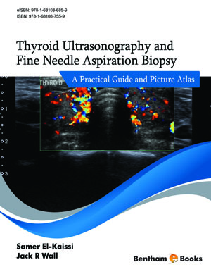
Thyroid Ultrasonography and Fine Needle Aspiration Biopsy: A Practical Guide and Picture Atlas is a concise and visual reference for diagnostic ultrasonography and needle biopsy of the human thyroid gland. The book provides the reader essential information on how to 1) distinguish between normal and abnormal thyroid sonograms, 2) differentiate low suspicion for malignancy thyroid nodules from sonographically high suspicion nodules, 3) evaluate cervical lymph nodes and parathyroid glands, and 4) examine post-thyroidectomy patients with differentiated thyroid cancer. The reader is also introduced to the different thyroid nodule risk stratification systems in ultrasound imaging, when and how to perform thyroid fine needle aspiration biopsies, and the use of percutaneous ethanol injections for cystic thyroid nodules. Key Features: -11 chapters which begin with an introduction to thyroid ultrasound and progressively explain relevant diagnostic imaging and biopsy procedures for different thyroid diseases (including thyroid cancer and autoimmune diseases) -multiple tables and figures which summarize and highlight important points -more than 60 ultrasound images which illustrate various ultrasound signs and artefacts from patients -a summary of the current standards for the evaluation and clinical management of thyroid nodules based on clinical practice guidelines -a detailed list of references, abbreviations and symbols The textbook is an essential reference for both practicing and training endocrine surgeons, endocrinologists, radiologists, cytopathologists, sonographers as well as any health care worker with an interest in managing thyroid and parathyroid diseases in their daily practice.


