Introduction: Cardiac Disease in the Pandemic Era: Teaching an Old Dog New Tricks?
Page: 1-5 (5)
Author: Ozgur KARCIOGLU*
DOI: 10.2174/9781681088204121010002
PDF Price: $15
Abstract
Nowadays, cardiac diseases, both developed de novo and acute exacerbations of chronic conditions, remain the most prominent death cause for the middle-aged and elderly, mostly in the developed, industrialized countries.
Since the end of 2019, COVID-19 pandemics have changed our lifestyles fundamentally, and maybe we will never find a way to return to the world of 2019. This catastrophic change had its impact on almost every aspect of our lives, including how we will manage cardiac arrest patients, how to perform perform cardiopulmonary resuscitation (CPR), ACLS, etc. A net effect is that protecting ourselves will take priority (more than before) in all procedures we pursue. Thus we can conclude that new generations should incorporate self-protecting behavior and techniques to benefit the patients in the most fruitful ways.
High-quality CPR cardiopulmonary resuscitation is among the most prominent issues to save humanity from the high burden of cardiac events. Relatively novel techniques such as mechanized devices for CPR, extracorporeal membrane oxygenation (ECMO), and therapeutic temperature management promise the highest possible solution to improve survival rates, in conjunction with urgent coronary angiography with revascularization.
Pandemics can be overcome not by the heroic behaviors of a few people but by the solidarity of society. The medical community should find the best solutions to help those in need with cardiac diseases even in pandemic conditions since this pandemic will not go away like magic. The aim of this book is to support patients and their next of kin, as well as health care workers, those who have dedicated themselves to healthy well-being with their relentless endeavor.
Cardiovascular Disease and COVID-19
Page: 6-18 (13)
Author: Ozgur KARCIOGLU*
DOI: 10.2174/9781681088204121010003
PDF Price: $15
Abstract
Cardiovascular disease (CVD) has long been the leading cause of global morbidity and mortality. However, with the COVID-19 pandemic, which has been the focus of attention all over the world since the end of 2019, this issue has gained different importance. The presence of CVD leads to more severe COVID-19 and an increased probability of mortality. In addition, both CVD and COVID-19 pave the way to myocardial injury, which also boosts the morbidity and death toll. Another point is the possible deprivation of usual healthcare received by cardiac patients (CVD and others) because of the shifted emphasis of the hospital and prehospital medical services on COVID-19. As the public can foresee that the pandemic will not disappear rapidly soon, healthcare organization faces a challenge to be redesigned radically. The objective of this chapter is to analyze CVD, myocardial injury, and other cardiac diseases resulting from COVID-19 itself, together with the impact of the pandemics on the usual healthcare of cardiac patients.
Myocardial Damage, Myocarditis, and COVID-19
Page: 19-30 (12)
Author: Ozgur KARCIOGLU*
DOI: 10.2174/9781681088204121010004
PDF Price: $15
Abstract
For centuries, complications of cardiovascular disease (CVD) have been documented as the prominent cause of mortality and morbidity worldwide. CVD and pandemic disease precipitate different kinds of damage in the myocardium, which also contribute to the death rates in the vulnerable population.
Almost all viral infections, including COVID-19 and influenza species, have the potential to inflict damage to the myocardial tissue, which also contributes to the severity of the disease itself. For instance, COVID-19 can trigger multiple organ failures with remarkable end results on cardiovascular functions. Its damage to the CVD like myocarditis, myocardial injury, de novo heart failure, acute coronary syndromes including STEMI, and various kinds of fatal and non-fatal dysrhythmias can be mentioned. Although most cases with myocarditis are asymptomatic or exhibit a mild course, it can precipitate acute heart failure and fatal respiratory failure.
Clinicians should be alert in patients with signs and symptoms compatible with myocarditis, pericarditis and/or endocarditis in the pandemic period and routine care because expedient diagnosis and management can prevent adverse outcomes in selected cases.
Coagulopathies, Prothrombotic State, Thromboembolism, Bleeding, and COVID-19
Page: 31-40 (10)
Author: Ozgur KARCIOGLU*
DOI: 10.2174/9781681088204121010005
PDF Price: $15
Abstract
COVID-19 is known to trigger a prothrombotic state, causing thromboses and thromboembolic events (TTEE) in patients with COVID-19. Both bleeding and thrombosis can result in significant morbidity in COVID-19. The entity paves the way to arterial TTEE (i.e., stroke and/or extremity ischemia) as well as small vessel thrombosis, which are commonly recorded at autopsy in the pulmonary vasculature. Elevated D-dimer is associated with a higher risk for TTEE, hemorrhage, critical illness, and mortality. Likewise, levels of fibrinogen, ferritin, procalcitonin are also higher in patients with thrombosis. There is also a propensity to develop pulmonary thromboembolism (PTE) in cases with COVID-19. Treatment with anticoagulant prophylaxis (i.e., heparin and/or aspirin) is recommended in many researches, but robust evidence is still warranted to draw firm conclusions on the benefit-to-harm ratio of the agents in most patients.
Chest Pain and Acute Coronary Syndromes (ACS)
Page: 41-107 (67)
Author: Ozgur KARCIOGLU*
DOI: 10.2174/9781681088204121010006
PDF Price: $15
Abstract
Acute coronary syndromes (ACS), especially acute myocardial infarction (AMI), is the leading cause of death in the world. These represent damage to the cardiac myocytes in the setting of acute cessation of blood supply. Chest pain is a common presentation in patients with AMI; however, there are multiple non-cardiac causes of chest pain. The diagnosis cannot always be made based on the initial presentation. The emergent evaluation of a patient with probable ACS includes a careful assessment of history, risk factors and presenting signs and symptoms, de novo ECG abnormalities, and workup of cardiac troponins. Validated risk scores, such as HEART, TIMI, and GRACE, can be helpful in predicting outcomes and the likelihood of ACS in a patient with chest pain. ECG should be performed within 10 minutes of presentation. ST elevation MI (STEMI) is diagnosed with elevated ST segments in two consecutive leads on ECG. Likewise, elevated levels of cardiac troponins in the first hours of presentation are mostly a prerequisite for diagnosis.
Although cardiac catheterization is viewed as the standard diagnostic modality for coronary artery disease, exercise testing, stress studies, echocardiography, and coronary computed tomography angiography (CCTA) may be important adjuncts. Cardiac catheterization laboratory (CCL), coronary care units, EDs, EMS, and primary care institutions need to cooperate in unison to produce the best results for public health.
This chapter gives a brief outline of the diagnosis and management of ACS in the pandemic period.
Heart Failure and Acute Pulmonary Edema (APEd)
Page: 108-136 (29)
Author: Ozgur KARCIOGLU*
DOI: 10.2174/9781681088204121010007
PDF Price: $15
Abstract
Heart failure (HF) is a complex syndrome in which the cardiac output cannot meet the demand, i.e., metabolic needs of the tissues and reflect the impairment of the heart's pump function. This condition is also referred to as congestive heart failure (CHF) as it is mostly associated with fluid retention.
The four main factors that determine the pump function of the left ventricle, which are contractility (contractility), preload, afterload and heart rate.
Accepted guidelines divided patients with HF into three groups according to their left ventricular ejection fraction (EF). The group with a EF below 40% continues to be known as a “low/reduced EF” (HF-REF), and a group of 50% and above remains “preserved EF” (HF-PEF), while a group of 40–49% is at the border (mid-range), thus it was named mildly reduced EF” (HF-MREF). The incidence of HF-PEF increases with age. The majority of cases in the elderly is due to HF-PEF. Acute decompensated HF is a deadly cause of cardiac dysfunction that can present with acute respiratory distress. There are many different causes of APEd, though cardiogenic pulmonary edema is usually a result of acutely elevated cardiac filling pressures. Clinical findings develop as a result of impaired perfusion and/or venous distension, with resultant surge in pressure. The patient mostly present with progressive symptoms of HF or acutely appeared signs of left-sided decompensation.
Patients who are diagnosed with HF for the first time and who is admitted with APEd should be hospitalized and treated accordingly. HF develops in 10 to 20% of AMI cases. Since this group has a high mortality, it must be identified and treated. The main objective of the treatment in the Acute Left HF is to provide the respiratory and cardiovascular stability as soon as possible. The main goal is to “dry” the lungs, not just throwing off water.
COVID-19 pneumonia and respiratory distress can masquerade APEd in the pandemic period. Most “typical” radiological findings including ground-glass opacities are common in both entities. It is very frequent that a clinician mixes up the two entities, especially misinterpret APEd as COVID-19, because the outbreak affects so many people that every physician is conditioned to see the viral pneumonia. Therefore, educational resources should stress on how to implement correct differential diagnosis of cardiopulmonary entities including AHF/APEd in the pandemics in both hospital and outpatient conditions. This chapter provides a general overview of the diagnosis and management of HF and APEd with a special emphasis on the acute presentation in the pandemic era.
Acute Pulmonary Embolism (APE)
Page: 137-153 (17)
Author: Ozgur KARCIOGLU*
DOI: 10.2174/9781681088204121010008
PDF Price: $15
Abstract
Acute pulmonary embolism (APE) is one of the diseases posing immense death rates and a great burden to public health. APE defines a blood clot or other substance in the deep leg/calf vein that traverses through the right heart and blocks the pulmonary arterial bloodflow (venous thromboembolism, VTE). The severity of the signs and symptoms of APE depends on the size of the thrombus and location of the occlusion, together with the previous reserves of the individual. A presentation template that will fit all cases cannot be put forward. “Massive” or hemodynamically unstable PE has a high death rate despite contemporary management. Healthcare personnel should be alerted to recognize untreated patient with high probability for APE in the ED and primary care institutions. Treatment should be expedient and aggressive in accord with the patient’s instability. Systemic or catheter-mediated thrombolysis, anticoagulation and other approaches should be contemplated immediately after general supportive measures.
This chapter delineates diagnostic dilemmas, distinctive properties and management principles of APE in the emergency setting. Also, challenges brought into scene with COVID-19 pandemics is discussed.
Hypertension and Aortic Diseases in The Pandemic Era
Page: 154-168 (15)
Author: Ozgur KARCIOGLU*
DOI: 10.2174/9781681088204121010009
PDF Price: $15
Abstract
The term hypertension (HT) is a chronic condition that leads to damage to target organs if untreated expediently. On the other hand, a hypertensive emergency refers to an acute elevation in blood pressure (BP) with evidence of end-organ injury, while hypertensive urgency defines acute BP elevation without progressive target organ dysfunction. Hypertensive emergencies comprise pulmonary edema/left ventricular failure, coronary syndromes, neurological deficits/intracranial hemorrhage, acute kidney injury, retinal hemorrhages, dissecting aortic aneurysm, and eclampsia.
The BP needs to be decreased expediently in the management of hypertensive emergencies. In the rest of the cases, the BP should be reduced in a gradual manner to preclude dangerously reduced cerebral perfusion pressure.
HT is also linked to COVID-19 as a comorbidity linked to a severe clinical course of the infection. On the other hand, dissecting aortic aneurysm (DAA) is most commonly seen after the age of 50 in hypertensive men who smoke. Most emergent aortic diseases appear to be a complication of HT and represents a major threat to public health. Clinicians should be alerted to recognize untreated patient with HT and aortic catastrophes in the emergency setting and primary care institutions.
Aortic Diseases: Abdominal Aortic Aneurysm (AAA) and Dissecting Aortic Aneurysm (DAA)
Page: 169-185 (17)
Author: Ozgur KARCIOGLU*
DOI: 10.2174/9781681088204121010010
PDF Price: $15
Abstract
Aneurysmal dilation is most common in the aorta, distal to the kidney vessels and proximal the iliac artery bifurcation. It is much more frequent in males than in females. It most commonly develops in middle aged and geriatric patients, patients with chronic HT, atherosclerosis, smoking history, and those with a genetic propensity for AAA, although none of this is an absolute rule.
The width of the aorta varies depending on the race, body area, gender and age, and the average aortic diameter is between 2.5 and 3.7 cm in general. Aortic diameter measuring 50% more (1.5 times) than expected is considered an aneurysm. If the diameter of the aorta is > 5 cm, the possibility of rupture increases and requires surgical intervention. In the abdominal aorta, which is generally located infrarenal,> 30 mm for both sexes is described as AAA.
In recent years, the term “Acute Aortic Syndrome” has also been used for all aortic emergencies. Signs and symptoms of AAA varies with the patient’s physiologic reserves, age and the extent of the disease with resultant organ damage (Table 1).
Supraventricular Arrhythmias and Their Management in the Emergency Setting: PSVT and AF
Page: 186-214 (29)
Author: Ozgur KARCIOGLU*
DOI: 10.2174/9781681088204121010011
PDF Price: $15
Abstract
Supraventricular tachycardia (SVT) is a type of tachyarrhythmia with a narrow QRS complex and regular rhythm (heart rate >100 bpm). These patients are often symptomatic and present to the emergency department (ED) in acute attacks called paroxysmal SVT (PSVT). Most SVTs are regular rhythms. It starts suddenly with the reentry mechanism in the majority of patients. 60% of the patients have reentry with Atrioventricular (AV) node, and 20% have reentry via bypass pathways. Coronary artery disease, anginal chest pain and dyspnea occur in patients due to tachycardia. Heart failure and pulmonary edema may occur with left ventricular dysfunction. Vagal maneuvers and adenosine appear to be the treatments of choice for termination of stable SVT.
Agents Used in the Treatment of Arrhythmias and Advanced Cardiovascular Life Support
Page: 215-238 (24)
Author: Ozgur KARCIOGLU*
DOI: 10.2174/9781681088204121010012
PDF Price: $15
Abstract
Advanced Cardiovascular Life Support (ACLS) guidelines recommend certain drugs for hemodynamic stabilization, prevention of collapse, stabilization of a perfusing rhythm, improving peripheral resistance and cardiac output, and restoration of organ perfusion. It is known that no antiarrhythmic agent increases the percentage of patients discharged with good neurological status. For this reason, the commencement of medications and establishing vascular access should not delay high-quality CPR.
ACLS guidelines recommend drug adrenaline in the asystole algorithm and in those with cardiac arrest due to ventricular fibrillation (VF). For pulseless electrical activity (PEA)-related cardiac arrest, adrenaline and, in some cases, sodium bicarbonate is recommended. The drugs used in VF and pulseless VT (PVT) apart from adrenaline are vasopressin, amiodarone, lidocaine, esmolol, magnesium, and procainamide in selected situations. This chapter provides a brief outline of arrhythmias commonly encountered in routine clinical practice, together with principles of ACLS and indications and usage of resuscitative agents employed in these situations.
Electrotherapies: Emergency Defibrillation, Cardioversion, and Transcutaneous Pacing
Page: 239-260 (22)
Author: Ozgur KARCIOGLU*
DOI: 10.2174/9781681088204121010013
PDF Price: $15
Abstract
Emergency cardioversion and defibrillation are life-saving procedures that exert direct electric current to the heart through the chest wall in order to terminate lethal tachyarrhythmias. Early defibrillation is life-saving in the survival of adult patients who develop sudden cardiac arrest. In the defibrillation process, myocardial cells are depolarized, and VF is terminated by delivering a certain amount of direct current to the heart, passing through the chest wall. Proper timing and accurate performance of these procedures have a vital role in both survival and recovery postresuscitation neurological functions without sequelae. Return of spontaneous circulation (ROSC) rates in defibrillation performed without losing time (within 20-30 seconds) can be up to 100% following the occurrence of these lethal rhythms. While cardioversion is performed in pulsating contraction rhythms, defibrillation is an electrical stimulation procedure applied in rhythms that do not generate pulses. In the cardioversion, synchronous energy is exerted onto the QRS complex to convert the rhythm into a sinus rhythm.
When there are signs of instability in rhythms with a pulse, emergency cardioversion (ECV) can be preferred over all other treatments if it is known to have acute onset (less than 48 hours) in atrial rhythm disorders, Transcutaneous pacing (TCP) is a recommended practice for temporary stabilization and invasive techniques such as transvenous pacing (TVP) should be attempted for longer pacing requirements. This chapter gives a brief outline on the outstanding features of electrotherapies (i.e., ECV; defibrillation; TCP, TVP) both in case of life-threatening dysrhythmias and also in urgent non-lethal situations.
Introduction
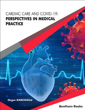
Cardiac Care and COVID-19: Perspectives in Medical Practice is an accessible reference on diagnoses and treatment modalities for cardiac diseases in general, and emergency cardiac conditions to be more specific, with respect to the current COVID-19 pandemic. Chapters in the book give updated descriptions of common problems in emergency medicine and cardiovascular disease. Each chapter is dedicated to a specific cardiovascular disease and explains management principles, diagnostic procedures and therapy. Examples of medical cases have also been used to highlight complex issues to give a concrete understanding of the cardiac care in COVID-19 patients to the medical practitioner, whether they are involved in critical care or in outpatient clinics. Key Features: - Clinical guidelines for critical care and cardiovascular management of COVID-19 patients - Topic-based information about cardiovascular diseases - Covers a range of cardiovascular problems including myocarditis, arrhythmias, chest pain, acute coronary syndrome - Information on pulmonary embolism and associated problems - Reader friendly presentation - Case-based examples for explaining concepts The range of topics combined with the simple presentation make this an essential reference for healthcare workers in emergency medicine, cardiology and nursing. General physicians interested in the cardiovascular impact of COVID-19 will also benefit from the information provided in the book.


