Book Volume 2
Editorial: Biomarkers to Biosensors - Technologies and Applications to Improve the Drug Discovery Process
Page: i-iii (3)
Author: Gary W. Caldwell, Atta ur-Rahman, Michael R. D`Andrea and M. Iqbal Choudhary
DOI: 10.2174/97816080520041060201000i
Strategies of Biomarker Discovery for Drug Development
Page: 5-22 (18)
Author: Xiang Jian Lou, S. M. Belkowski, James M. Dixon, Brenda Hertzog, Dan Horowitz, Sergey I. Ilyin, Danielle Lawrence and D. Polkovit
DOI: 10.2174/978160805200410602010005
Abstract
Drug development is a long and costly process. Although the length of drug development may vary depending on the target class, attrition is the main contributor to the financial burden. Therefore, various biological markers (biomarkers) are needed to determine compound efficacy and dosing. Biomarkers provide comprehensive information about the molecular network of the target and lead compound in vivo. Such information has the potential of shortening time of development and reducing costs by facilitating the decision of what lead compound should move forward at early development stages. This review article focuses on the strategies for biomarker discovery by giving readers a practical guideline to approaches that are used for the discovery, validation and use of biomarkers to accelerate the process of drug discovery.
Protein and Antibody Microarrays: Clues Towards Biomarker Discover
Page: 23-33 (11)
Author: Kazue Usui-Aoki, Motoki Kyo, Makoto Kawai, Masatoshi Murakami, Kazuhide Imai, Kiyo Shimada and Hisashi Koga
DOI: 10.2174/978160805200410602010023
Abstract
The recent advancement of proteomics technologies has provided us a variety of approaches for protein-expression profiling. Among these approaches, protein and antibody microarrays are promising new ones for biomarker discovery. Although at present they have several limitations with respect to sample preparation, sensitivity, specificity, and so on, protein and antibody microarrays will no doubt become a standard adjunctive method in the actual clinical scene. With this in mind, we have been establishing a novel system for antibody microarray in which surface plasmon resonance (SPR) technology is utilized for the signal detection. Up to 400 real-time antibodytarget bindings could be measured simultaneously within a single hour. Although SPR is assumed to be an expedient technology for protein and antibody microarrays, here we describe its advantages and disadvantaged compared to other detection technologies. This review focuses on the technological aspects of these two methods and a discussion of their clinical usefulness. We further emphasize the interpretation of the protein and antibody microarray results in combination with the results of DNA microarray and intracellular pathways mainly constructed from data on protein-protein interaction.
The Use of Biomarkers to Detect Cervical Neoplasia and to Diagnose High-Grade Cervical Disease
Page: 35-54 (20)
Author: Douglas P. Malinowski
DOI: 10.2174/978160805200410602010035
PDF Price: $15
Abstract
The detection of cervical carcinoma and its malignant precursors is currently accomplished using the Pap smear in conjunction with testing for the presence of human papillomavirus (HPV). This screening approach is successful at identifying patients with cervical disease with a very high sensitivity, but with limited specificity. In order to improve the accuracy of cervical disease detection and diagnosis, a number of approaches have been employed to incorporate molecular diagnostics into the testing procedure. Two investigational approaches to identify biomarkers have been employed: (1) the use of specific biomarkers based upon known keratinocyte response to HPV infection; and (2) the use of genome-wide expression profiles to identify new genes whose expression is altered in response to HPV infection and the transformation process. Both approaches have identified biomarkers that appear suitable for the detection of the mild dysplasia precursors of disease, the malignant precursors of moderate-severe dysplasia and cervical carcinoma. Some biomarkers are suitable for the detection of HPV infected cells displaying mild dysplasia while others are more specific to moderate-severe dysplasia and carcinoma (disease-specific markers). The disease-specific markers appear to be over-expressed in high-grade cervical disease and represent aberrant entry of the infected cells into the S-phase of the cell cycle. These markers appear promising in molecular diagnostic applications to detect malignant cells in both histology and cervical cytology specimens. An emerging diagnostic paradigm will be discussed where HPV DNA analysis represents measurements of transient infection and risk for future disease; analysis of HPV oncogene transcripts distinguishes active versus transient infection; and detection of aberrant S-phase induction represents a measure of active disease.
New Developments in the Field of Protein and Metabolism Assays Aimed at Drug Discovery Processes
Page: 55-86 (32)
Author: Kothandaraman Narasimhan, Ponnusamy Sukumar and Mahesh Choolani
DOI: 10.2174/978160805200410602010055
PDF Price: $15
Abstract
The advent of new high-throughput proteomic and metabolic assays using mass spectrometry (MS) has significantly benefited drug discovery process. This process is accelerated with the miniaturization of detection devises (nanotechnology and biosensors) to carry out rapid and effective screening. Using proteomics (protein arrays) it is possible to globally investigate the molecular basis of disease, drug action leading to drug development. Similarly metabolic assays for nutritional status, expanded neonatal screening through tandem MS could swift through several thousand data points associated with a particular disease (either proteins/peptides or metabolites) in a short span of few minutes and come up with highly sensitive and accurate diagnosis. Nanobiotechnology raises fascinating possibilities for new analytical array based assays (receptor-ligand binding, DNA-DNA hybridization, or both) and microanalytic separations, each of which will be mentioned here with respect to their ability to affect the drug discovery processes. Molecular profiling by DNA microarray technology has made significant contributions to the understanding of molecular targets for diseases such as cancer. One of the challenges is how to efficiently utilize the accumulated research data to develop new diagnostic and/or prognostic markers and therapeutic targets. Proteomics based disease therapeutic research involves high-throughput protein structure determination (e.g. structural biology of protein tyrosine kinases (PTKs). These processes involve conventional antibody based arrays as well as targets identified using structural biology for narrowing down targets for drug delivery. Drug discovery processes aimed at generating inhibitors for the treatment of malignancies are believed to be dependent on the gain of function of specific PTKs. Current research include Src as a target for pharmaceutical intervention, JAK kinases in leukemias/lymphomas, and phosphoproteomics. The following areas mentioned above will form the key focus of this chapter.
Proteomic Screening for Novel Therapeutic Targets in Kidney Diseases
Page: 87-102 (16)
Author: Visith Thongboonker
DOI: 10.2174/978160805200410602010087
PDF Price: $15
Abstract
The high-throughput capability of proteomics allows simultaneous examination of numerous proteins and makes a global analysis of proteins in cell, tissue, organ or biofluid possible. This strength of proteomics has been extensively applied to examine altered proteins caused by various diseases. Some of these altered proteins may particularly be important for the disease progression and/or complications. Thus, information on these altered proteins is valuable for better understanding of the pathogenesis and pathophysiology of medical diseases. Additionally, functional analysis of such proteins may lead to identification of novel therapeutic targets and development of new drugs for improving therapeutic outcome as well as for preventing serious complications. This review focuses mainly on applications of renal and urinary proteomics to define novel therapeutic targets in kidney diseases. Several recent studies on various kidney diseases have successfully identified altered renal and urinary proteins, some of which may potentially be the novel therapeutic targets. Urinary proteome profiling has also been applied to biomarker discovery that will be useful for clinical diagnostics, prognosis, prediction of treatment response, and development of personalized medicine. Finally, potential roles of proteomics for drug design and discovery are discussed.
Aptamer-Based Technologies as New Tools for Proteomics in Diagnosis and Therapy
Page: 103-119 (17)
Author: Vittorio de Franciscis and Laura Cerchia
DOI: 10.2174/978160805200410602010103
PDF Price: $15
Abstract
Proteomics has provided a tool to define protein profile of a specific cell or tissue and to associate protein expression levels and post-translational modifications with disease states therefore developing innovative technologies for measurement of protein levels has become a major challenge of the last few years. Specific nucleic acid-based compounds, named aptamers, have been shown as high-affinity ligands and potential antagonists of disease-associated proteins. Aptamers, isolated from combinatorial libraries by an iterative in vitro selection process, discriminate between closely related targets thus representing a valid alternative to antibodies or other bio-mimetic receptors, for the development of biosensors and other bio-analytical methods. Moreover they can be easily stabilized by chemical modifications for in vivo applications and numerous examples have shown that stabilized aptamers against extracellular targets such as growth factors, receptors, hormones or coagulation factors are very effective inhibitors of the corresponding protein function. By integrating the aptamer-based biosensor development with the maturing technology for in vitro selection of anti-protein aptamers results in the highthroughput production of proteome chips. Furthermore, aptamer arrays and biosensors will reveal the most effective tools for the detection of biomolecular interactions and the identification of protein targets, particularly with regard to those not detectable by known receptors like enzymes or antibodies. We will review here the main and innovative methods based on the use of aptamers as biosensors for protein detection that, in alternative or combined to the classical proteomic approaches, could reveal suitable for both diagnostic and therapeutic purposes.
Recent Developments in Proteomics: Mass Spectroscopy and Protein Arrays
Page: 121-150 (30)
Author: Aarohi Kulkarni and Mala Rao
DOI: 10.2174/978160805200410602010121
PDF Price: $15
Abstract
Proteomics has been gaining increasing attention in order to understand the relevance of the genomic information available through high throughput DNA sequencing and microarray techniques. Proteomics can be viewed as an experimental approach to explain the information contained in the genomic sequences in terms of the structure, function and control of biological processes and pathways. It thus systematically analyses the proteins expressed in the cell. It is well known that several modifications of proteins like posttranslational modifications and splicing are not visible at the DNA level but alter the functions of the proteins involved greatly. These can be envisaged at the protein level wherein the functions can be assigned. Coherent strategies and technologies are required to elucidate protein expression, interactions and functions. The most well established approach to the identification of proteins and their separation is the 2-D PAGE. It allows the resolution and identification of proteins from a wide variety of sources without understanding their functions. There are limitations to this approach, which lacks sensitivity and its inability to separate effectively hydrophobic membrane proteins. The technique however remains the leader methodology for delivery of protein expression and for recording change in protein structure. Recently mass spectrometry has become the method of choice for the identification and characterization of proteins after their purification. In the current methods of mass spectroscopy the protein is identified from fragments and as such does not yield information about posttranslational modifications. Its application is still at a developing stage and in future it will be one of the leader techniques for depicting protein structure. Proteomics has recently seen advancement in the form of microarray technique. Microarray based analysis is a high throughput technology, which can be performed at a dynamic range and also has the added advantage of performing functional proteomics. Its ease of operation and ability to control key parameters such as temperature, pH and cofactor concentrations makes it suitable to automation. This review will essentially focus on the different mass spectroscopy approaches being used to identify and characterize proteins from diverse sources and the developments in protein array technology.
NMR Spectroscopy Based Metabonomics: Current Technology and Applications
Page: 151-173 (23)
Author: Clare A. Daykin and Florian Wulfert
DOI: 10.2174/978160805200410602010151
PDF Price: $15
Abstract
Metabonomics has, in the past decade demonstrated enormous potential in furthering the understanding of disease processes, toxicological processes, phenotypic outcome of gene expression and biomarker discovery. However, implementation of metabonomic methodology requires the development of generic, rapid, advanced analytical tools to comprehensively profile biosample (biofluids and tissues) metabolites. 1H NMR spectroscopy is arguably the most powerful tool for profiling of biosamples with inherent advantages over other analytical techniques e.g.: 1) lack of requirement for extensive sample preparation; 2) non-destructive; 3) non-equilibrium perturbing; 4) small (down to 1 ml) sample volumes and 5) quantitative and qualitative information are available from the same data set. Whilst conventional `target analysis` approaches may apply more sensitive analytical methods than 1H NMR spectroscopy for detection of low metabolite levels in biological materials, the target analysis approach involves consideration of critical biochemical pathways and pre-selection of the metabolites of interest. The subjective selection of a battery of biochemical methods is then required which is a necessary, but complex and time-consuming process and if an inappropriate or restricted range of biochemical methods or parameters are chosen, important metabolic disturbances may be over-looked. On the other hand, the use of 1H NMR spectroscopy to follow biochemical responses does not require such a pre-selection of metabolites and allows subsequent multicomponent analysis, without bias imposed by the experimenters expectations, hence it has been demonstrated in a wealth of publications that NMR spectra of biosamples are extraordinarily rich in information on endogenous biochemical processes in both good health and disease.
This review will aim to discuss technical developments relevant to the field, commonly used data handling and data analysis strategies and the use of metabonomics as a tool to facilitate our understanding of health status at the metabolic level.
NMR-Based Metabonomics of Urine from An Exploratory Study of Ciprofibrate in Healthy Volunteers and Patients with Type 2 Diabete
Page: 175-191 (17)
Author: Gregory C. Leo, Ewoud J. van Hoogdalem and Martijn B.A. van Doorn
DOI: 10.2174/978160805200410602010175
PDF Price: $15
Abstract
This report presents the results from the NMR-based metabonomics of urine collected as part of a multiple dose, double-blind, randomized, placebo controlled parallel group exploratory study of the pharmacodynamics of ciprofibrate in healthy volunteers and patients with type 2 Diabetes Mellitus. Using supervised statistical analysis it was possible to distinguish the various treatment groups (healthy and diabetic: placebo-treated or drug-treated) when males and females were analyzed separately.
Chromatography Mass Spectrometry Based Metabonomic Analytical Methods
Page: 193-209 (17)
Author: Gary W. Caldwell, Wensheng Lang, Gregory C. Leo, John A. Masucci, William Jones and Andrew Mahan
DOI: 10.2174/978160805200410602010193
PDF Price: $15
Abstract
The concept of a system biology approach to understanding biological functions such as the development and the progression of chronic diseases and drug-induced toxicity in humans has generated widespread interest in the pharmaceutical community. Techniques including genomics, transcriptomics, proteomics, peptidomics, lipidomics and metabolomics are being integrated not only to understand the entire biological system but also to discover novel biomarkers. Analytical “Xomics” assays are playing a major role in the development of these methods. We will review some of these analytical techniques and specifically focus on chromatography mass spectrometry based metabonomic strategies applied to diverse areas such as fermentation, bacteria, plants and animals.
OWLS - A Versatile Technique for Drug Discovery
Page: 211-223 (13)
Author: Jeremy J. Ramsden
DOI: 10.2174/978160805200410602010211
PDF Price: $15
Abstract
Optical waveguide lightmode spectroscopy (OWLS) is forecast to become a key measurement technology in drug design and discovery. This review focuses on what the technology is, why it is superior to existing technologies and how it may be expected to develop for drug discovery applications.
Cell-Based Biosensors in Proteomic Analysis
Page: 225-239 (15)
Author: Spiridon E. Kintzios
DOI: 10.2174/978160805200410602010225
PDF Price: $15
Abstract
In recent years there has been a rapid increase in the number of diagnostic applications based on biosensors, including live, intact cells, tissues, organs or whole organisms. Whole cells provide multipurpose catalysts, particularly in processes that require the participation of a number of enzymes in sequence. However, the sensitivity and reliability of these sensors is often limited by the signal transduction mechanisms and by non-specific interferences, due both to analyte and environmental variations.
In similar fashion to DNA and protein microarrays, which deliver multiplex detection via the high-density spatial arrangement of molecular recognition elements, arrays of cells at high-density can form the basis of cell-based sensors with extremely high-throughput capability. The expression of receptors of interest within these arrays could yield cell-based sensors with defined specificities. In addition, transfected cell microarrays composed of highdensity arrays of mammalian cells expressing defined genes, could be the basis for future high-throughput cell-based protein sensing platforms. Such cellular arrays could be used for the detection of molecular interactions in functional proteomics in vitro, to the testing of proteins in functional studies in living cells. Microarrays with ordered cell arrangements of GFP-producing or luminescent bacteria may be used as an integral part of future biosensors. Recent and representative applications in this direction include (i) the profiling of antibody specificities and protein interactions with genetically engineered human immune cells, (ii) cells containing surface antibodies, specific to antigens of different pathogens and (iii) cell proliferation/metabolism sensors dedicated to screening for drug candidates and drug kinetic analysis.
Current Approaches in Natural BiopolymerNanoparticle Hybrid Functional Materials: From Drug Delivery to Bio-Detection Applications
Page: 241-258 (18)
Author: Roberta Brayner
DOI: 10.2174/978160805200410602010241
PDF Price: $15
Abstract
This review is focused on current approaches emerging at the intersection of hybrid nanomaterials research and biotechnology. This interdisciplinary field of chemistry and biology is also associated with physical and chemical properties of organic and inorganic nanoparticles, as well as to DNA, proteins and polysaccharide studies.
Spectroscopic Analysis of Cell Physiology and Function
Page: 259-272 (14)
Author: Mark Riley, Iram Mondaca Fernandez and Pierre Lucas
DOI: 10.2174/978160805200410602010259
PDF Price: $15
Abstract
Spectroscopic methods including infrared and Raman techniques have tremendous promise for providing rapid, non-invasive information on the impact of pharmaceuticals and toxicants on cells and tissues. Spectroscopy is not a new field; however these methods have only recently been applied to study the physiology and function of cells and tissue. The infrared spectrum (2,500-25,000 nm) of a cell or tissue can supply information about the fundamental vibrational modes of functional groups existing in biological molecules thus permitting quantification of material composition and in some cases can be connected to cell function. Spectroscopy permits observation of changes within the cell occurring at the molecular level and is a rapid, nondestructive, and reagent-free measurement.
Infrared and Raman spectroscopy have been applied to evaluate the biochemical composition of mammalian cells or to discriminate between cancerous and healthy cells. While discriminations can be made between stages of the cell cycle due to alterations in spectral signatures of nucleic acids, only very small differences are apparent between distinct subcellular structures. Analyses of separate cell fractions, isolated by sucrose density gradient centrifugation, present the most significant spectral differences in the C-H (carbon and hydrogen) stretching region (2800-3000 cm-1), at the ester carbonyl stretching band (1737 cm-1), and in the PO2 - stretching region (1089 and 1242 cm-1).
This manuscript provides a critical review of these methods, including their potential use and pitfalls for drug screening applications. Significant innovations have been made over recent years in equipment, experimental approaches, and in analysis methods.
Encapsulated Biomolecules Using Sol-Gel Reaction for High-Throughput Screening
Page: 273-294 (22)
Author: Kumiko Sakai-Kato, Masaru Kato, Naoko Utsunomiya-Tate and Toshimasa Toyo`oka
DOI: 10.2174/978160805200410602010273
PDF Price: $15
Abstract
Recently, the sol-gel encapsulation method has attracted much attention for the development of desirable protein-doped matrices as biosensors. Proteins are entrapped into a porous, silica matrix that is formed via a low-temperature sol-gel reaction. The encapsulated proteins can retain their structure and biological activity for a prolonged period. This sol-gel encapsulation method allowed the reuse of expensive protein reagents multiple times. Furthermore, the encapsulation method often improved the stability of the immobilized proteins. Based on these reasons, this technology has been used in various fields, and is expected to contribute to the effectiveness of analytical systems and the application to high-throughput screening systems. In this review, we introduce various studies in which biomolecules were immobilized on capillary-, microchip-, and microarray-based analytical systems using the sol-gel reaction. The interactions of the immobilized biomolecules and analytes were detected using UV, fluorescence, mass spectrometry, or electrochemical detection. On the other hand, many researchers are studying the sol-gel processing to improve the biocompatibility of the sol-gel derived materials using new biocompatible silane precursors and processing methods. The microstructure of the silica matrix was also investigated using various analytical systems. We also review some reports that described the fundamental aspects of the sol-gel reaction.
Modeling of Environmentally Sensitive Hydrogels for Drug Delivery: An Overview and Recent Developments
Page: 295-331 (37)
Author: Hua Li, Rongmo Luo and K.Y. Lam
DOI: 10.2174/978160805200410602010295
PDF Price: $15
Abstract
A critical review of mathematical modeling for simulation of environmentally sensitive hydrogels is presented for application of drug delivery. The review demonstrates that there have been large numbers of published studies on the model development, although the majority of investigations in the research area of the drug delivery are experimental-based. Therefore, a systematical review of mathematical modeling of environmentally sensitive hydrogels is necessarily provided through comprehensive assessment on several critical developments of mathematical models to simulate the hydrogels for mechanisms of drug release. The present review classifies the developed models into the fundamental and empirical/semi-empirical groups and also discusses the properties and performance of the hydrogels as drug delivery system in kinetics and equilibrium.
Polyelectrolyte Nanocapsules - Promising Progress in Development of New Drugs and Therapies
Page: 333-348 (16)
Author: Silke Krol, Alessandra Gliozzi and Alberto Diaspro
DOI: 10.2174/978160805200410602010333
PDF Price: $15
Abstract
The most promising tool for future applications in the field of science as well as in medicine is the use of nanobiotechnologies. Especially self-assembly systems with tailored properties on a nanometer level fulfill the requirements to nano-organized systems in a satisfactorily manner. Hence the development of so-called nanocapsules prepared by means of Layer-by-Layer technique was a great progress on the way to individual drug delivery systems or nano-sized bioreactors. The preparation of hollow shells for drug delivery use requires polyelectrolytes as well as a charged core that are not cytotoxic. According to this purpose CaCO3 crystals with different shapes were introduced as removable template for capsules with changeable permeability as a result of pH variations. Due to the low toxic potential of the core it could be valuable for applications in human body.
Furthermore the nano-organized shells are suitable as coating of living cells or artificial tissue. With this “second” cell wall it is possible to target the encapsulated material to predefined organs, and to prevent immune response. Moreover one can choose between the breakage of the coverage using the capsule only as targeted carrier or the production of proteins inside the remaining shell. The requirements for this application are polyelectrolytes that are not toxic to the tissue of the transplantation site as well as to the coated cells.
Contributors
Page: 349-352 (4)
Author: Atta ur-Rahman, Gary W. Caldwell, M. Iqbal Choudhary and Michael R. D’Andrea
DOI: 10.2174/978160805200410602010349
Subject Index to Volume 2
Page: 353-365 (13)
Author: Atta ur-Rahman, Gary W. Caldwell, M. Iqbal Choudhary and Michael R. D’Andrea
DOI: 10.2174/978160805200410602010353
Introduction
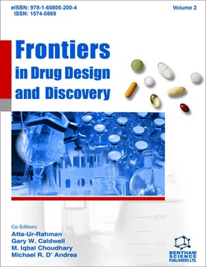
Frontiers in Drug Design and Discovery is a book series devoted to publishing the latest and the most important advances in drug design and discovery. Eminent scientists write contributions on all areas of rational drug design and drug discovery including medicinal chemistry, in-silico drug design, combinatorial chemistry, high-throughput screening, drug targets, and structure-activity relationships. The book series should prove to be of interest to all the pharmaceutical scientists involved in research in drug design and discovery. Each volume is devoted to the major advances in drug design and discovery. The book series is essential reading to all scientists involved in drug design and discovery who wish to keep abreast of rapid and important developments in the field. The book series is available in print, online and CD editions.


