Contributors
Page: iii-v (3)
Author: Kunlin Jin and Guo-Yuan Yang
DOI: 10.2174/978160805001710901010iii
Calpain Modulation of Programmed Cell Death Pathways Following Cerebral Ischemia
Page: 1-8 (8)
Author: P. S. Vosler and J. Chen
DOI: 10.2174/978160805001710901010001
PDF Price: $15
Abstract
Cerebral ischemia causes a massive insult resulting in the eventual death of ischemia-affected neurons. Historically, research efforts have focused on the canonical cell death signaling pathways examining the activation of both caspase-dependent and -independent mechanisms to execute neuronal death due to ischemia. Recently, however, there is evidence that the calcium-activated protease calpain is able to mediate both neuronal death pathways. This chapter briefly outlines the intrinsic and extrinsic caspase-dependent pathways and the caspase-independent pathways. This is followed by a discussion of the role of calpain in abrogating the caspase-dependent pathway and instigating the caspase-independent pathway. Greater understanding of how neurons actuate delayed neuronal death will potentially lead to the development of viable therapeutics to diminish the negative neurological sequelae caused by cerebral ischemia.
Calcium-permeable Ion Channels and Ischemic Brain Injury
Page: 9-17 (9)
Author: Theresa A. Lusardi, Xiangping Chu and Zhi-Gang Xiong
DOI: 10.2174/978160805001710901010009
PDF Price: $15
Abstract
Stroke, or brain ischemia, is a leading cause of morbidity and mortality worldwide. It is also a leading cause for long-term disabilities. Although enormous progresses have been made in recent years towards defining the responses of brain to ischemia, thrombolysis remains the only effective treatment for stroke patients. Unfortunately, thrombolitics have limited success and a serious side effect of intracerebral hemorrhage. It has been well recognized for many years that excessive Ca2+ accumulation in neurons is essential for neuronal injury associated with brain ischemia. However, the exact mechanism(s) and pathway(s) underlying the toxic Ca2+ loading remain elusive. The objective of this chapter is to discuss the role of several major Ca2+-permeable cation channels, including NMDA-receptor-gated channels, TRPM7 channels, and acid sensing channels, in glutamate-dependent and independent Ca2+ toxicity associated with brain ischemia.
The Protective Effect of Ischemic Postconditioning Against Ischemic Brain Injury
Page: 18-25 (8)
Author: Heng Zhao
DOI: 10.2174/978160805001710901010018
PDF Price: $15
Abstract
Ischemic postconditioning is an emerging concept for stroke treatment. It refers to a series of mechanical interruptions of reperfusion after ischemia, preventing ischemia/reperfusion injury in both myocardial and cerebral infarction. This review article reveals that the earliest study about ischemic postconditioning was performed more than 50 years ago in the research field of myocardial ischemia, and only thrilled in recent 5 years, and it was shifted from myocardial ischemia to cerebral ischemia only 2 to 3 years ago. The protective effect of postconditioning has been studied in focal and global ischemia in vivo, and in slice or primary neuronal cultures in vitro. In addition, protective parameters of postconditioning in various ischemic models are discussed. Thereafter, this article provides insights on postconditioning's protective mechanisms associated with reperfusion injury, the Akt, MAPK and PKC cell signaling pathways, suggesting that postconditioning attenuates free radical generation, that the Akt pathway contribute to its protection, and that the MAPK and PKC pathways are closely associated with its protection.
Neovascularization Following Cerebral Ischemia
Page: 26-37 (12)
Author: Rodney Allanigue Gabriel and Guo-Yuan Yang
DOI: 10.2174/978160805001710901010026
PDF Price: $15
Abstract
Neovascularization is the generation of new blood vessels and is made possible either through vasculogenesis, arteriogenesis, or angiogenesis. This process is far from simple as a plethora of growth factors, cytokines and chemokines, and various cell types are required to interact in a collaborative manner in order to initiate and maintain neovasculature. Because neovascularization process occurs following ischemia or traumatic injury, promoting neovascularization is a potential therapeutic approach to these insults. Exogenous regulation of blood vessel formation is therapeutic when it produces functional and stable capillaries, in which newly proliferating microvessels minimally increase blood-brain-barrier permeability and produce adequate regional cerebral blood flow. To review this issue of cerebral neovascularization, we discuss: 1) important angiogenic growth factors, cytokines, extracellular matrix proteins, and cell types involved in brain angiogenesis; 2) involvement of inflammation in cerebral neovascularization; 3) stem cells play a role in cerebral neovascularization; 4) neovascularization following cerebral ischemia in animal model or in clinical cases; and finally 5) neovascularization as a therapeutic target for cerebral ischemic injury.
Role of Matrix Metalloproteinases After Stroke: From Basic Research to Clinical Impact
Page: 38-45 (8)
Author: Anna Rosell, Eng H. Lo and Xiaoying Wang
DOI: 10.2174/978160805001710901010038
PDF Price: $15
Abstract
Matrix metalloproteinases (MMPs) comprise a family of zinc endopeptidases that play major roles in the physiology and pathology of the mammalian central nervous system (CNS). These proteinases are evolutionarily conserved as modulators of extracellular matrix during CNS development. After acute tissue injury such as that which occurs after stroke, MMPs become dysregulated and subsequently mediate acute neurovascular disruption and parenchymal destruction. Animal studies have demonstrated their participation in breakdown of neurovascular matrix and blood-brain barrier disruption with edema and/or hemorrhage. Moreover, perturbation of extracellular homeostasis triggered by MMPs may underlie processes responsible for the hemorrhagic complications of thrombolytic stroke therapy. Conversely, biphasic roles for some MMPs have been established since emerging data also suggest that some aspects of MMP activity during the delayed neuroinflammatory response may contribute to remodeling and stroke recovery.
Blood Brain Barrier Dysfunction and the Endothelin System in Cerebral Ischemia
Page: 46-51 (6)
Author: Samuel W. Cramer, Lin Li and Dandan Sun
DOI: 10.2174/978160805001710901010046
PDF Price: $15
Abstract
The blood brain barrier (BBB) is the central homeostatic controller of the brain environment and plays an important role in disease. Disruption of normal BBB functionality is a significant event in the pathogenesis of cerebral ischemia. Therefore, strategies that attenuate BBB disruption during cerebral ischemia and reperfusion represent viable therapeutic approaches capable of decrease the severity of ischemic injury, including reducing the risk of hemorrhage and edema formation. The endothelin (ET) system comprises three peptides (ET-1, ET-2, and ET-3) and two receptor sub-types (ETA and ETB) [1,2]. This system is involved in a diverse array of physiological process. The ET system is also plays an integral role in BBB dysfunction through the modulation of ion transporters, water channels, and the recruitment of various cellular mediators of the inflammatory response. Because of this involvement, the ET system represents a possible therapeutic target for the treatment of cerebral ischemia. This review examines the interplay between the BBB and the ET system in cerebral ischemia.
Research Progress of Hypothermia: Selective Intra-arterial Infusion and Regional Brain Cooling in Acute Stroke Therapy
Page: 52-62 (11)
Author: Yuchuan Ding and Justin Charles Clark
DOI: 10.2174/978160805001710901010052
PDF Price: $15
Abstract
In the United States, stroke is the 3rd leading cause of death, behind diseases of the heart and cancer, and the number 1 cause of disability. Basic research on stroke has been extensive, but clinically effective therapies are still lacking. Recombinant tissue plasminogen activator (tPA) is the only drug approved by the Food and Drug Administration (FDA) for selected patients (3%) with ischemic stroke. The benefit of this thrombolytic therapy is largely limited by it's selection criteria and its side effects. An area of stroke research that holds much promise is regional brain cooling. The neuroprotective effect of hypothermia has long been recognized. Currently used whole body cooling for brain injury treatment from stroke was abandoned because of management problems, severe side effects and delayed onset of cerebral hypothermia. Recent studies in a rat stroke model have utilized a unique technique to infuse the microvasculature in the ischemic territory prior to reperfusion with cold saline, leading to a improved outcomes in this animal model. Translating the research done with combined intra-arterial revascularization and local brain cooling to the clinical realm could advance the treatment of stroke beyond the levels achieved by current therapies.
Stem Cell Transplantation and Cerebral Ischemia
Page: 63-73 (11)
Author: Christine L. Keogh, Shan Ping Yu and Ling Wei
DOI: 10.2174/978160805001710901010063
PDF Price: $15
Abstract
Stroke is caused by a partial or full blockage of the blood supply to certain part of the brain and afflicts both young and old. Stroke can be either hemorrhagic or ischemic in nature and can also result from a permanent blockage or a transient occlusion of one or several arteries. The transient stroke may additionally involve reperfusion injury. Currently, the only effective means of stroke treatment is early administration of tissue plasminogen activator (tPA), which has a small window of opportunity (within 3 hours after the onset of stroke). Thus development of new therapies especially the delayed treatments many hours and even days after stroke is very much needed. One treatment approach that has recently garnered a lot of attention is stem cell transplantation. Much of neuroscience research focuses on the analysis and characterization of both the endogenous response of stem cell proliferation and migration following an ischemic insult as well as the effects of exogenous stem cell transplantation. Studies have identified endogenous stem cell niches in the adult and the neonatal rodent brain and many groups have tried to harness the ability of the host to regenerate damaged tissue following stroke. This chapter will briefly review endogenous stem cell experimentation and cover recent advancements in exogenous stem cell transplantation.
Post-ischemic Neurogenesis and Brain Repair: Growth Factors and Cytokines
Page: 74-82 (9)
Author: Yi-Ping Yan, Raghu Vemuganti and Robert J. Dempsey
DOI: 10.2174/978160805001710901010074
PDF Price: $15
Abstract
Stroke is the major cause of neurologic death and disability in the adult population. The persistence of neurogenesis in the adult mammalian brain has brought hope that endogenous neural progenitors may be a potential source to repair the damaged brain after cerebral ischemia. In the adult mammalian brain, neurogenesis takes place in specific regions, including the sub-ventricular zone (SVZ) of the lateral ventricle, the sub-granular zone (SGZ) of the dentate gyrus (DG) in the hippocampus and the posterior peri-ventricle (PPV) dorsal to the hippocampus. Accumulating evidence indicates that cerebral ischemia stimulates neurogenesis in adult brain. Numerous attempts in the past to better understand the complicated mechanisms of ischemia-induced neurogenesis have revealed several crucial processes such as proliferation of neural progenitors, migration and differentiation of newly-generated cells, as well as functional incorporation of these cells in the injured brain. However, the molecular mechanisms regulating these steps are far from clearly understood. This review summarizes the role of growth factors and cytokines in the regulation of neurogenesis following cerebral ischemia. The functional significance of post-ischemic neurogenesis, as well as the future research directions to achieve improved functional recovery after stroke by enhancing this endogenous process of brain repair are also discussed.
Brain Aging, Neurogenesis and Experimental Stroke
Page: 83-89 (7)
Author: Kunlin Jin
DOI: 10.2174/978160805001710901010083
PDF Price: $15
Abstract
Aging is associated with a striking increase in the incidence of stroke and neurodegenerative diseases, both of which are major causes of disability among those aged 70 years and older in the United States. Despite progress in understanding the molecular mechanisms of neuronal cell death in these diseases, widely effective treatments remain elusive. Adult endogenous neural stem cells hold great promise for brain repair because of their unique location within the central nervous system, their potential to proliferate and to differentiate into all major neural lineages, and their ability to incorporate functionally into existing neuronal circuitry after stroke. Nevertheless, the ability to exploit these cells for therapeutic purposes is hampered by the lack of knowledge about the biological behaviors of neural stem cells in the adult brain, and the cellular and molecular signals that control the generation of a functional neuron from adult neural stem cells after stroke, particularly in the aged brain. Therefore, it is essential to better understand the biological behaviors of neural stem cells and how neural stem cells are regulated after stroke in the aged brain, since stroke affects mainly the aged population. In this regard, brain aging and neurogenesis after focal cerebral ischemia are reviewed in this chapter.
Erythropoietin and Ischemic Brain Remodeling
Page: 90-93 (4)
Author: Zheng Gang Zhang and Michael Chopp
DOI: 10.2174/978160805001710901010090
PDF Price: $15
Abstract
Erythropoietin (EPO) is a hematopoietic cytokine and has the neuroprotective effect for treatment of acute ischemic stroke. Emerging data indicate that EPO also plays an important role in brain remodeling after stroke. This chapter reviews the effect of EPO on neurogenesis and angiogenesis and signaling pathways that mediate EPO-enhanced coupling of neurovascular niche in the ischemic brain.
Potential MRI Methodologies and Treatment of Stroke
Page: 94-99 (6)
Author: Quan Jiang
DOI: 10.2174/978160805001710901010094
PDF Price: $15
Abstract
Magnetic resonance imaging (MRI) has shown great potential in early detection and characterization of stroke injury. MRI methodologies such as diffusion and perfusion MRI have been successfully applied to both experimental stroke and clinical studies, covering a broad range of questions raised in the early treatment of stroke. However, potential MRI methodologies related to critical issues in the treatment of stroke have not been well reviewed. In the current review, we present an overview of possible MRI methodologies addressing the critical issues related to treatment of stroke, including MRI measurements for predicting and detecting hemorrhagic transformation, as well as staging ischemic tissue.
The Use of a Global Statistical Approach for the Design and Data Analysis of Clinical Trials with Multiple Primary Outcomes
Page: 100-108 (9)
Author: Peng Huang, Robert F. Woolson and Ann-Charlotte Granholm
DOI: 10.2174/978160805001710901010100
PDF Price: $15
Abstract
Determining whether one treatment is preferred over others is a major goal of many clinical studies but can be complicated by the situation when no single outcome is sufficient to make the judgment. We present a useful global statistical test technique and the corresponding global treatment effect (GTE) measure for assessing treatment's global preference when multiple outcomes are evaluated together. Applications of these techniques in clinical trial design and data analysis are illustrated.
Introduction
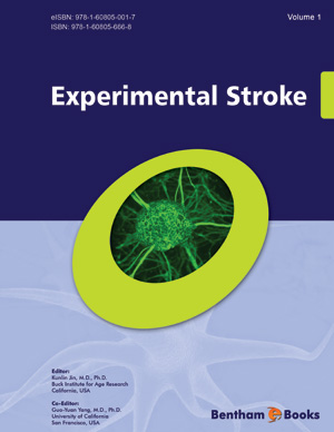
This eBook compiles the efforts of 20 experts in the field to review the latest advances in experimental stroke, with its strong emphasis on neurogenesis, angiogenesis and neuroprotection after cerebral ischemic stroke. It also provides current data for pharmacologic therapy, including anti-Inflammation, for stroke. Several chapters address cell death and apoptotsis after stroke as well as its replacement strategies by stem cells. Other chapters deal with the relation between stroke and neuroimmunology, BBB disruption and electrophysiological changes. Also covered is magnetic resonance imaging in ischemic brain. In a variety of preclinical models of stroke, pre-conditional and post-conditional stroke models will be discussed.


