Preface
Page: ii- (1)
Author: Anna Strunecka and Russell L. Blaylock
DOI: 10.2174/9781608051960110010002
List of Contributors
Page: iii-iii (1)
Author: Anna Strunecka, Russell L. Blaylock, Mark A. Hyman and Ivo Paclt
DOI: 10.2174/978160805196011001010iii
Autism Spectrum Disorders: Clinical Aspects
Page: 1-16 (16)
Author: Ivo Paclt and Anna Strunecka
DOI: 10.2174/978160805196011001010001
PDF Price: $30
Abstract
Autism spectrum disorders (ASD) are a group of related neurodevelopmental disorders, which includes autism (autistic disorder), Asperger syndrome, Rett syndrome, pervasive developmental disorder-not-otherwise specified (PDD-NOS), and childhood disintegrative disorder (CDD). This chapter provides the review of recent knowledge about clinical symptoms and criteria for diagnosis of heterogeneous symptoms of ASD. An alarming increase in the prevalence of ASD is of great concern to practicing pediatricians and psychiatrists. Some people attribute the increases over time in the frequency of ASD to factors such as new administrative classifications, changing diagnostic criteria, and heightened awareness. It is evident, that no single factor or a simple explanation can account for the increase. ASD are highly genetic and multifactorial, with many risk factors acting together. There is no therapy of the core symptoms of ASD at present. Several studies suggest that 50-75% of children with ASD are using complementary alternative medicine. Families and clinicians need access to theoretical and clinical evidence to assist them in the choice of therapies.
The Cerebellum in Autism Spectrum Disorders
Page: 17-31 (15)
Author: Russell L. Blaylock
DOI: 10.2174/978160805196011001010017
PDF Price: $30
Abstract
The cerebellum is the most commonly affected part of the brain in autistic spectrum disorders (ASDs). The histopathological changes strongly indicate selected damage to particular cell groups and lobules of the cerebellum rather than diffuse injury. A number of studies have shown injury and abnormal development of the vermis of the cerebellum, with a predominance of neuronal loss among Purkinje cells and granule cells. In addition, one see abnormal pathway development indicating intrauterine damage or damage occurring during the early postnatal period. Several studies have shown abnormalities of glutamate receptors (GluRs) of various kinds, including metabotropic GluRs (mGluRs). In this chapter, I review the histopathologic findings within the ASD cerebellum and demonstrate evidence for immunoexcitotoxicity affecting cerebellar neurodevelopment as well as evidence for early neurodegeneration. Newer studies have shown that the cerebellum may have significant cognitive and higher cortical functions, either by way of its connections to prefrontal-limbic areas or more indirect pathways.
Dysregulation of Glutamatergic Neurotransmission in Autism Spectrum Disorders
Page: 32-46 (15)
Author: Anna Strunecka
DOI: 10.2174/978160805196011001010032
PDF Price: $30
Abstract
Despite the great number of observations being made concerning cellular and molecular dysfunctions associated with autism spectrum disorders (ASD), an integrative and unifying mechanism to explain the heterogeneous symptoms and etiology of ASD has not been proposed in the major scientific literature. We offer the explanation of potential etiology of ASD as dysregulation of glutamatergic neurotransmission with underlying interactions between chronic microglial activation and the excitotoxic cascade playing the central role. This chapter summarizes current knowledge of the structural and functional diversity of glutamate receptors (GluRs) and excitatory amino acid transporters. Recent research of the autism genome also supports the view that abnormalities in genes connected with glutamate neurotransmission and disturbed regulation of glutamate pathways may be directly involved in ASD. We further suggest that the increasing prevalence of ASD during the last decades might reflect the synergistic action of an increased burden of new excitotoxic factors. In this chapter we discuss the effects of dietary excitatory amino acids, mainly glutamate and aspartate, which could exacerbate the pathological and clinical symptoms of ASD. The mechanism of excitotoxicity is the topic of the next chapter.
Immunoexcitotoxicity as a Central Mechanism of Autism Spectrum Disorders
Page: 47-72 (26)
Author: Russell L. Blaylock
DOI: 10.2174/978160805196011001010047
PDF Price: $30
Abstract
Autism has undergone a tremendous amount of study, and a number of often seemingly unconnected disorders have been disclosed. Yet, despite an enormous amount of study, no central mechanism to explain the causation of this syndrome or why it affects only a subset of children has come forth. In this chapter I propose such a central mechanism that explains a great number of biochemical, histological, neurodevelopmental and systemic dysfunctions, as well as behavioral findings in autism spectrum disorders (ASD). Since the discovery of excitotoxicity by Olney in 1968, neuroscientists have determined that not only is glutamate a neurotransmitter, but it is the most abundant neurotransmitter in the brain, exceeding the more traditional neurotransmitters combined. Recent studies have also shown that glutamatergic receptors (GluRs) interact with other receptors, not only neurotransmitters, but also immune receptors, in a way that can alter their sensitivity. Chronic brain inflammation is known to dramatically enhance the sensitivity of N-methyl-D-aspartic acid (NMDA) and α- amino-3-hydroxy-5-methyl-4-isoxazole propionic acid (AMPA) type GluRs and interfere with glutamate removal from the extraneuronal space, where it can trigger excitotoxicity and abnormal synaptic and dendritic physiology over a prolonged period. Importantly, neuroscience studies have clearly shown that sequential systemic immune stimulation can not only activate the brain ’ s immune system, microglia and astrocytes, but that there occurs an amplified response to both subsequent stimulation, either systemic or within the CNS. The ASD child is exposed to such sequential immune stimulation via a growing number of vaccines, recurrent infections, chemical toxins and persistent viral infections.
Immune Dysfunction in Autism Spectrum Disorders
Page: 73-81 (9)
Author: Russell L. Blaylock
DOI: 10.2174/978160805196011001010073
PDF Price: $30
Abstract
A great number of studies have been done examining immune function in children with autism spectrum disorders (ASD). Most of these studies have demonstrated immune dysfunction, especially involving cellular immunity. Important to the immunoexcitotoxicity hypothesis is the finding that macrophages and lymphocytes from ASD children have been shown to demonstrate an amplified release of pro-inflammatory cytokines with stimulation, especially in those having gastrointestinal (GI) symptoms. Because of the intimate connection between the gut and brain, hyperimmune responses from the gut, vial vagal afferents, can rapidly activate brain microglia, leading to an exaggerated innate immune response within the brain. It has also been shown that ASD children often react to food peptides, such as gliadin, gluten and casein as well as a number of bacterial and fungal antigens, all of which can exaggerate immunoexcitotoxicity. The finding of cross-reacting food antigen with brain components also indicates the presence of bystander damage and would trigger immunoexcitotoxicity as well.
Gastrointestinal Disorders and Autism Spectrum Disorders: A Causal Link or a Secondary Consequence?
Page: 82-99 (18)
Author: Anna Strunecka
DOI: 10.2174/978160805196011001010082
PDF Price: $30
Abstract
Growing evidence confirms that up to 95% of autistic children suffer with the dysfunctions of the gastrointestinal (GI) system. We discuss the cellular and molecular mechanisms underlying these disturbances. Some researchers, physicians, and health care professionals suggest that beneficial effects of dietary intervention on behavior and cognition of some autistic children indicate a functional relationship between the GI tract (GIT) and the CNS pathology of ASD. A possible genetic cause for the association of autism and GI disease is discussed. GI disorders are not included in diagnostic criteria for ASD. Clinical and practical experiences provide the support for association between inflammatory bowel disease and ASD.
Biochemical Changes in ASD
Page: 100-120 (21)
Author: Anna Strunecka
DOI: 10.2174/978160805196011001010100
PDF Price: $30
Abstract
Metabolic dysfunctions have not been extensively studied in ASD despite the fact that chronic biochemical imbalance is often a primary factor in the development of several neurological diseases. Substantial percentages of autistic patients display peripheral markers of mitochondrial energy metabolism dysfunction, such as elevated lactate and alanine levels in blood and serum carnitine deficiency. We assess the reported biochemical changes in the blood and evidence based on the exploration of brain imaging studies. Even though alterations in mitochondrial and cellular energy metabolism are not specific for ASD, they indicate the potential ethiopathological events. Evidence from several laboratories similarly indicates that biomarkers of oxidative stress may be increased in some autistic children. One of the best documented biochemical changes in ASD is a decrease in cellular glutathione (GSH) levels, a major intracellular antioxidant, and an increase in oxidized glutathione (GSSG). Alterations in methionine -homocysteine cycle have been studied in details in ASD. Significant changes in transmethylation and transsulfuration metabolites in plasma from autistic children were reported. The new finding indicates a significant decrease in methylation capacity and redox potential. Metabolic and mitochondrial defects may have toxic effects on brain cells, causing neuronal loss and altered modulation of neurotransmission systems. The observations of biochemical changes thus further support that the antioxidant therapy and supplementation with some vitamins could prevent and restore the energy metabolism of individuals with ASD. This chapter brings evidence of the impact of observed biochemical changes in ASD for potential amelioration of ASD symptoms and for evidence-based therapy.
Searching the Role of Mercury in Autism Spectrum Disorders
Page: 121-147 (27)
Author: Anna Strunecka and Russell L. Blaylock
DOI: 10.2174/978160805196011001010121
PDF Price: $30
Abstract
Mercury is a ubiquitous environmental toxin that causes a wide range of adverse health effects in humans. The population is now exposed to mercury mostly from seafood consumption, dental amalgam, vaccines, and certain pharmaceuticals. Exposure to mercury can cause neurological, immune, sensory, motor, and behavioral dysfunctions similar to traits defining or associated with autism spectrum disorders (ASD). Because of an observed increase in autism in the last decades, which parallels cumulative mercury exposure, it was proposed that autism may be, in part, caused by mercury. The autism-mercury hypothesis has generated much interest and controversy. Thimerosal, a preservative added to many vaccines, has become one of the potential culprits in the pathogenesis of ASD. This chapter shows that the overwhelming preponderance of the evidence favors acceptance of the hypothesis that mercury exposure is capable of causing some ASD if exposure occurs at critical developmental periods. Special attention is paid to forms of mercury of current public health concern, which include vapors of metallic mercury from dental amalgam, methylmercury in edible tissues of fish and whales, and ethylmercury from thimerosal added to certain vaccines. Mercury in all of its forms is toxic to the fetus and children, and efforts should be made to reduce exposure to pregnant women and children as well as the general population. Moreover, emerging evidence supports the theory that some ASD may result from a combination of genetic/biochemical susceptibility and synergistic action of mercury with other excitotoxic contaminants from the environment.
Fluoride and Aluminum: Possible Risk Factors in Etiopathogenesis of Autism Spectrum Disorders
Page: 148-161 (14)
Author: Anna Strunecka and Russell L. Blaylock
DOI: 10.2174/978160805196011001010148
PDF Price: $30
Abstract
Fluoride and aluminum ions (Al3+) are considered as new ecotoxicological factors. While aluminum has been involved among the possible culprits of autism spectrum disorders (ASD), fluoride is rarely considered. Al3+ is non-essential for all forms of life and serves no known biological role. Fluoride and Al3+ can elicit impairment of homeostasis, growth, development, cognition, and behavior. Several symptoms induced by Al3+ and/or fluoride overload can be seen in ASD. Several laboratory studies demonstrate that many effects primarily attributed to fluoride are caused by synergistic action of fluoride plus Al3+. In water solutions, Al3+ forms in the presence of fluoride, water soluble aluminofluoride complexes (AlFx). AlFx has been widely used as an analogue of phosphate groups to study heterotrimeric G proteins involvement. AlFx affects numerous receptors and signaling systems. It is evident that the long-term intake of low amounts of fluoride and Al3+ can evoke receptor malfunctions. The synergistic interactions of fluoride plus Al3+ may thus evoke several histological, neurological, biochemical, and behavioral symptoms of ASD. This chapter brings evidence that AlFx represents a hidden potential danger for pathogenesis of ASD.
The Role of Melatonin in Etiopathogenesis and Therapy of Autism Spectrum Disorders
Page: 162-172 (11)
Author: Anna Strunecka, Russell L. Blaylock, Mark A. Hyman and Ivo Paclt
DOI: 10.2174/978160805196011001010162
PDF Price: $30
Abstract
Pineal melatonin, an endogenous signal of darkness, is believed to be an important regulator of circadian and seasonal rhythms. The changing melatonin levels serve as hands of a bio-clock and dates of the biocalendar in vertebrates including humans. Circulating melatonin regulates and influences the sleep wake cycle, sexual development, as well as various immune, endocrine, and metabolic functions. Initially it was thought to be produced exclusively by pineal gland. Subsequently, it was shown that melatonin is also produced in several other tissues. Substantial amounts of melatonin are found in the gut as well as the brain. Moreover, melatonin is one of the most powerful scavengers of free radicals. Sleep disorders and low melatonin levels are frequently observed in people with autism spectrum disorders (ASD). The role of abnormal melatonin biosynthesis in the gastrointestinal system relating to the development of ASD symptoms warrants further studies. The important multiple role of melatonin in prevention and amelioration of ASD symptoms is not fully recognized at present. Melatonin appears to be promising as one of the efficient and seemingly safe adjunctive treatments in children and adults with ASD.
The Search for Plausible Role of Oxytocin in Etiology and Therapy of Autism Spectrum Disorders
Page: 173-185 (13)
Author: Anna Strunecka, Russell L. Blaylock, Mark A. Hyman and Ivo Paclt
DOI: 10.2174/978160805196011001010173
PDF Price: $30
Abstract
The discoveries of novel sites of oxytocin receptor (OTR) expression in central nervous system have greatly expanded the classical biological roles of oxytocin (OT), which are stimulation of uterine smooth muscle contraction at parturition and milk ejection during lactation. Central actions of OT range from the modulation of the response on individual synapses to the establishment of complex social and bonding behaviors. While there are currently no animal models reflecting the broad range of the autism spectrum disorders (ASD) behavioral and neurological phenotypes, studies of vole pair bonding and sexual behavior provided important clues useful for understanding the neurobiological mechanisms of social behavior. It has been suggested that OT dysfunction might contribute to the development of social deficits in autism, a core symptom domain and potential target for intervention. The intranasal administration of OT has been reported to reduce repetitive behaviors in adult patients with ASD. The studies of the possible role of OT in ASD etiology have raised interest but also brought some contradictory findings. Though there are preliminary promising findings with OT intranasal delivery in autistic adults, further studies are needed to replicate these findings on a large scale. Studies concerning the safety of these potential treatments are needed as well.
Regulation of Cortisol Levels in Autistic Individuals and their Mothers
Page: 186-198 (13)
Author: Anna Strunecka, Russell L. Blaylock, Mark A. Hyman and Ivo Paclt
DOI: 10.2174/978160805196011001010186
PDF Price: $30
Abstract
The significant evidence has been collected that individuals with autism spectrum disorders (ASD) have disturbed regulation of the hypothalamic-pituitary-adrenal (HPA) axis. Disturbance in daily rhythm, the increased level of pituitary adrenocorticotropin hormone (ACTH), and the decreased concentration of cortisol in plasma and saliva has been found. The hypoactivity of HPA axis and decreased level of saliva cortisol has also been found in parents of autistic individuals. Adverse maternal stress during gestation is involved in abnormal behavior, mental and cognition disorders in offspring. Hippocampus is the principal target site for corticosteroids in the brain as it has the highest concentration of receptor sites for glucocorticoids. Several studies found that repeated prenatal glucocorticoid exposure has profound influences on HPA function both in animals and humans. Lower cortisol is seen in conditions of chronic stress and in social situations characterized by unstable social relationships. The presence of social support has been associated with decreased stress responsiveness and with attenuated free cortisol concentrations in saliva. Several studies document that oxytocin significantly reduced salivary cortisol levels. Findings from several laboratories, which investigated both autistic children and parents, further support the knowledge that mothers may share some metabolic characteristics with their autistic children and that there is the need to intervention of both - autistic children and their mothers. It has been suggested that paired-like homeodomain transcription factor 1(PITX1), a key regulator of hormones within the HPA axis, may be implicated in the etiology of autism. The role of 11.-hydroxysteroid dehydrogenase type 1 (11.-HSD1) in the pathogenesis of ASD is discussed.
Reproductive Hormones and Autism Spectrum Disorders
Page: 199-205 (7)
Author: Russell L. Blaylock
DOI: 10.2174/978160805196011001010199
PDF Price: $30
Abstract
There is some evidence that autistic individuals have elevated exposure to androgenic hormones, even female autistics, and that autistic behaviors may, in part, be explained by influences of testosterone on neurodevelopment and higher-order brain function. Some studies indicate early exposure to maternal levels of testosterone in utero may play a predominate role. Similarities of ASD disorders to childhood schizophrenia are strengthened by a similar relationship between high levels of androgen exposure early in life and this disorder. The fact that estrogens play a major role in neuroprotection may explain the male preponderance of ASD. Recent studies have shown that testosterone can enhance excitotoxicity and estrogen can reduce excitotoxicity. Rather than a direct sex hormonal effect on brain function, I propose that the sex hormones are playing a modulating role on immunoexcitotoxicity.
Addendum. Autism: Is It All in the Head?
Page: 206-216 (11)
Author: Mark A. Hyman
DOI: 10.2174/978160805196011001010206
PDF Price: $30
Abstract
Mark A. Hyman, MD, is Chairman of the Institute for Functional Medicine. Anna Strunecka asked him to provide his Editorial [1] as an addendum of this eBook. Autism is described as a hologram for chronic disease; an extreme manifestation of disruptions in normal biology that exist in varying degrees in most chronic illness. Autism is a complex, multi-system disorder rooted in a series of toxic, infectious, and allergic insults. Through the story of one boy, the author looked carefully at the few biological systems manifesting as the clinical features of autism: gut and immune dysfunction, nutritional deficiencies, toxicity and impaired detoxification, mitochondrial dysfunction and oxidative stress, and genetic polymorphisms that set the stage for biochemical train wrecks. In autistic children, the results of testing often reveal results that show deviations orders of magnitude higher than in other chronic illness, but nonetheless, the same patterns exist. The lessons learned from the dissection of the functional causes and mechanisms of autism can illuminate the path for whole system medicine and clinical research and the potential for it to address the global crisis of chronic disease.
Index
Page: 217-223 (7)
Author: Anna Strunecka, Russell L. Blaylock, Mark A. Hyman and Ivo Paclt
DOI: 10.2174/978160805196011001010217
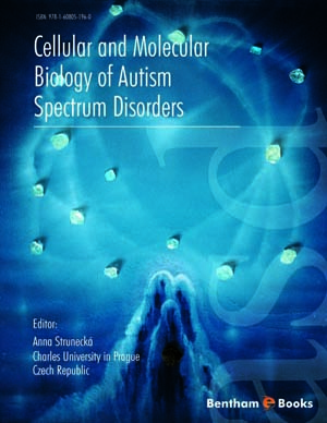
Introduction
Over the past several decades the incidence of autism spectrum disorders (ASD) has increased dramatically. The etiology of ASD remains an unsolved puzzle to scientists, physicians, pediatricians, psychiatrists, and pharmacologists. Our E-book will address what is presently known concerning the pathophysiology of ASD from a cellular and molecular perspective. Our explanation is based on the interaction between repetitive systemic immune stimulation with concomitant chronic brain activation of microglia, which leads to overstimulation of glutamate receptors and inflammatory cytokine receptors. Our E-book will explain, for the first time, the effects of immunoexcitotoxicity on the brain development, neurophysiology, and pathology. Our book will not only attempt to explain the finding in ASD, but will offer treatment proposals that address each of these mechanisms. It will also explain how previous, often successful treatment methods, may indeed operate through the immunoexcitotoxic mechanism.


