Foreword
Page: i-i (1)
Author: Guenter J. Krejs
DOI: 10.2174/9781681087870121010001
Preface
Page: ii-ii (1)
Author: Ioan Sporea and Alina Popescu
DOI: 10.2174/9781681087870121010002
List of Contributors
Page: iii-vi (4)
Author: Ioan Sporea and Alina Popescu
DOI: 10.2174/9781681087870121010003
What’s New in Extra-digestive Gastroesophageal Reflux Disease?
Page: 1-16 (16)
Author: Vasile-Liviu Drug* and Oana-Bogdana Bărboi
DOI: 10.2174/9781681087870121010004
Abstract
Gastroesophageal reflux disease (GERD) is a highly prevalent complex chronic condition. The most extensive prospective and multicenter cohort study conducted in Europe has estimated that one-third of the patients with GERD may exhibit extra-esophageal symptoms. The Montreal Consensus recognized chronic cough, chronic laryngitis, bronchial asthma and tooth erosions as extra-digestive manifestations of GERD. The experts also considered that manifestations such as recurrent otitis media, idiopathic pulmonary fibrosis, sinusitis or pharyngitis are likely to be associated with GERD. The traditional techniques used in the diagnosis of typical GERD are less useful for the diagnosis of extra-digestive GERD. No single testing methodology exists to definitively identify reflux as the etiology for the suspected extra-esophageal symptoms. The PPI trial is the first diagnostic but also a therapeutic step, while evaluation through esophageal impedance-pH monitoring currently represents the goldstandard for diagnosis. Despite extensive work, extra-digestive GERD remains incompletely understood.
Optical Diagnosis in Barrett Esophagus and Related Neoplazia
Page: 17-25 (9)
Author: Daniela E. Dobru*
DOI: 10.2174/9781681087870121010005
Abstract
The detection of high grade dysplasia and esophageal adenocarcinoma with improved survival rates is the aim of optical diagnosis in BE. Advanced imaging technologies improve the characterization of dysplastic BE by mucosal visualization and enhancement of the fine structural and microvascular details (mucosal and vascular pattern) and may guide targeted biopsies for the detection of dysplasia during surveillance of patients with previously non-dysplastic BE.
Eosinophilic Gastrointestinal Disorders
Page: 26-43 (18)
Author: Dan L. Dumitraşcu* and Andrei V. Pop
DOI: 10.2174/9781681087870121010006
Abstract
Eosinophilic infiltration of the gut occurs unusually and its clinical relevance was only recently recognized. The medical conditions with eosinophilic infiltration are commonly named eosinophilic gastrointestinal disorders [EGID]. EGID is described as a gastrointestinal tract disorder with functional and morphological abnormalities due to a dense infiltration of eosinophils in the gastrointestinal wall. The cause could be an allergic reaction due to varied allergens, food or the environment. EGID is including eosinophilic esophagitis [EoE], eosinophilic gastroenteritis [EGE], and eosinophilic colitis [EC]. EGIDs pathophysiology is not yet fully understood, but histopathology is characterized by degranulation and an excessive number of eosinophils. A role in the pathophysiology of EGIDs is played by a hypersensitive reaction. Diagnosing EGIDs is quite challenging. It can be described as a combination of eosinophilic invasion of one or more organs from the GI tract with non-specific GI symptoms. The gold standard for EGIDs diagnosis is the histology of gastrointestinal mucosal biopsy, an overabundance of eosinophils being the principal diagnostic criterion without a known cause. The treatment for EGID is not well defined yet, because of the limited prospective controlled studies performed. The treatment is an empiric one and is administrated according to the severity of the symptoms and it is represented by diet, corticosteroids, and steroid agents.
Postoperative Digestive Complications of Bariatric Surgery
Page: 44-55 (12)
Author: Andrada Seicean* and Radu Seicean
DOI: 10.2174/9781681087870121010007
Abstract
Digestive complications of bariatric surgery are quite rare, especially those which are sever in nature, with a lower rate aftersleeve gastrectomy compared to Rouxen-Y gastric bypass. This chapter discusses the bleeding, anastomotic leaks, stenosis and ulceration, gastroesophageal reflux, bowel transit dysfunction, gallstones and complications related to adjustable gastric banding and other after bariatric surgeries.
What is New in Gastro-Entero-Pancreatic Neuroendocrine Tumors
Page: 56-66 (11)
Author: Adrian Săftoiu* and Codruța Constantinescu
DOI: 10.2174/9781681087870121010008
Abstract
Gastroenteropancreatic neuroendocrine tumors (GEP-NETs) are a group of heterogeneous malignancies that can occur anywhere in the digestive system, with a growing incidence over the past decade. For proper diagnosis and management, the grading and histological diagnosis have been revised recently. Thus, the WHO grading criteria have been updated in 2017 as well as the TNM staging for pancreatic NETs in 2018. To establish a correct diagnosis, a multimodal approach is required, including various biomarkers, endoscopic tumor biopsy and tumor imaging. Over the past decades, improved diagnostic techniques including endoscopic ultrasound and somatostatin receptor fusion imaging have gained ground and have assisted treatment decision making. Regarding the treatment strategy, the management implies taking into account the tumor stage and degree of tumor differentiation, as well as tumour growth and spread. Novel therapies such as molecular-targeted agents, tryptophan hydroxylase inhibitor and peptide receptor radionuclide therapy were recently approved by FDA, improving the prognosis for advanced GEP-NETs.
Intestinal Microbiota and its Implications in Pathology
Page: 67-78 (12)
Author: Paul J. Porr*
DOI: 10.2174/9781681087870121010009
Abstract
The intestinal microbiota develops as a results of various genetic, nutritional and environmental factors, becoming very specific for each individual. It totalizes more than 100 trillions of bacteria with a piece of genetic information more than 100x greater than the human genome. The functions of the microbiota can be grouped into metabolic, protective and structural. The microbiota-derived metabolites signal to distant organs of the host, which enable the microbiota to connect to the brain, the immune and endocrine system, metabolism and other functions of the host. These microbiota-host communications are essential to maintain the vital functions and health of our organism. So, microbiota, in eubiosis and especially in dysbiosis, has multiple effects on the human organism. The therapeutic possibilities for this are the administration of nonabsorbable antibiotics, pre-, pro, syn- or symbiotics, as well as FMT, which is in principle a complex human probiotic. The most important digestive effects of microbiota are in Clostridium difficiledetermined pseudomembranous colitis, in IBS, IBD, diverticulitis, functional dyspepsia, and in different digestive cancers: gastric, colorectal, liver and pancreatic cancer. Alcoholic liver disease is also influenced by microbiota. The extra-digestive effects of microbiota are very complex. In some metabolic diseases, like obesity, NAFLD, atherosclerosis, dyslipidemias and T2D, special types of dysbiosis have important pathophysiologic implications. Microbiota has also implications in Alzheimer's disease, osteoporosis, CKD, different psychiatric disorders and some extra-digestive cancers. In conclusion, it may be stated that the intestinal microbiota has multiple effects, even in diseases that apparently have no relation with the intestinal flora.
Videocapsule Endoscopy
Page: 79-90 (12)
Author: Ciprian Brisc* and Timothy Kurniawan
DOI: 10.2174/9781681087870122010010
Abstract
Videocapsule endoscopy is a non-invasive and important innovation in diagnostic endoscopy. This technology was first launched in 2000 and it has been widely used by gastroenterologists worldwide. This method requires the patient to swallow a miniature high-resolution camera which will pass through the digestive tract, while transmitting images to the recorder in order to be evaluated. It has its main advantages which are the non-invasiveness, and the possibility to yield a diagnosis in severely ill patients who cannot support invasive endoscopy procedures, but it also has disadvantages which include the impossibility to perform a biopsy or other therapeutic procedures. Over the years, this method has been revolutionized by not only approaching the small bowel, but also the esophagus and the colon. This chapter will also discuss the application of the esophagus capsule as well as the colon capsule. There are multiple indications for which patients can be referred to videocapsule endoscopy. The most frequent cause of referral to capsule endoscopy is the obscure GI bleeding, but it may be used in detecting small intestine polyps or tumors, searching for the cause of iron deficiency anemia or reviewing the extension of Crohn’s disease. The main risk of this method is represented by retention which is also minimal.
Precision Medicine in Inflammatory Bowel Disease: Current Challenges
Page: 91-103 (13)
Author: Eugen Dumitru and Cristina Tocia*
DOI: 10.2174/9781681087870122010011
Abstract
Fibrosis in Crohn’s Disease - From Evolution to Treatment
Page: 104-113 (10)
Author: Adrian Goldiş*
DOI: 10.2174/9781681087870121010012
Abstract
One of the major complications of Crohn's disease is the development of fibrosis, this causes the intestine to lose its mobility. The most frequent intestinal “damage” occurrences are considered fibrosis, fistula, abscess, resected bowel. The Lemann index has been developed to describe the entire gut damage score in CD. It is summarizes the clinical, imaging, endoscopic, and surgical findings from all the segments of the digestive tract into one global score and provides a superior quantification of the severity of bowel, destruction. Chronic inflammation, hypertrophy of MP (muscularis propria) and smooth muscle hyperplasia of SM (submucosa) were the most valid histopathological features characterizing the intestinal stricture. Imaging methods such as MRI, CT or IUS can detect penetrating disease and intra-abdominal abscesses in different accuracy grades. Although the current imaging techniques were not able to determine the degree of fibrosis, MRI was preferred in the US for pelvic fistulae, abscesses or deep-seated fistulae. By decreasing MRTF and p38 MAPK activation and increasing autophagy in fibroblasts, local ROCK inhibition prevents and reverses intestinal fibrosis. Fibrosis is certainly reversible in animal models. The duration of treatment and toxicity are challenging for the time being.
The Quality of Life of Patients with Inflammatory Bowel Disease: A Continuous Challenge
Page: 114-122 (9)
Author: Mircea Diculescu and Tudor Stroie*
DOI: 10.2174/9781681087870121010013
Abstract
Inflammatory bowel diseases (IBDs) are chronic conditions of the gastrointestinal tract with a remitting and relapsing course and an unpredictable evolution. Patients affected by these diseases often have to deal with severe abdominal pain, diarrhea and loss of bowel control, fatigue, multiple surgeries and a wide range of extra-intestinal manifestations. Given these facts, the majority of them have a severely impaired health-related quality of life (HR QoL) and they are more prone to developing anxiety and depression. Even though early clinical trials didn’t show much interest in it, assessing the patients’ QoL has become, over time, one of the main endpoints of the clinical trials, thus more and more articles involving the patients’ QoL being published every year. Patients with active disease have a significantly lower HR QoL compared to those with inactive disease. Regarding the disease phenotype, especially when in remission, patients with Crohn’s disease tend to have lower QoL than those with ulcerative colitis. Anxiety and depression have a significant impact on the patients’ HR QoL. Another concern regarding the patients with IBD is the high rates of fatigue. Fatigue is a common symptom in many other inflammatory conditions like rheumatoid arthritis or multiple sclerosis, and leads to a significant impairment of the QoL and lowers work productivity. In spite of this, it is frequently underdiagnosed or overseen by physicians, and many times remains unexplored and untreated.
Advances in Colorectal Cancer Screening
Page: 123-133 (11)
Author: Eftimie Miuțescu and Bogdan Miuțescu*
DOI: 10.2174/9781681087870121010014
Abstract
Colorectal cancer (CRC) is the second most commonly diagnosed cancer in women and the third most commonly diagnosed cancer in men. There is a 5% lifetime risk of developing CRC in many regions and despite treatment, 45% of persons diagnosed with CRC die as a result of the disease. The development of molecular biology techniques and methods has allowed a thorough knowledge of the carcinogenicity process in the CCR. Currently, multiple guidelines are available that provide guidance to clinicians who refer patients to screening. Although colonoscopy is the preferred tool for detecting and diagnosing CCR, non-invasive stool-based tests are widely used. In this section we reviewed the most important studies that have been published regarding molecular biomarkers to identify new approaches, as well as metabolomics for identifying new biomarkers for colorectal cancer. Death occurring from colorectal cancer can be prevented by detecting cancer and precancerous lesions at an early stage. For achieving this goal, new screening tools are mandatory and research for better screening tests is needed.
New Guidelines on Post-polypectomy Colonoscopy Surveillance
Page: 134-144 (11)
Author: Simona Băţagă*
DOI: 10.2174/9781681087870121010015
Abstract
Colorectal cancer (CRC) remains a frequent tumor, in spite of the screening programs developed in most of the countries. It is well known that CRC is developing from polyps and that the polypectomy prevents the CRC and ultimately the death of the patient. One important debate is about the post polypectomy surveillance of the patients, in regard to the timing of the second colonoscopy after the baseline one. Appropriate intervals spare the patient from an unwanted colonoscopy, however, in the case of advanced lesions ensures no recurrence of the lesion. Last year, important guidelines were elaborated and revised by different societies. This chapter is summarizing the recent European, American and British guidelines which are mostly similar, with small exceptions. The updated guidelines are reducing the number of colonoscopies in patients with small adenoma and serrated polyps without dysplasia. The villous proportion of a polyp is not considered a risk factor. In the piece-meal resection is indicated a shorter period to reevaluate the patient to reduce the risk of incomplete resection. The present guidelines are decreasing the unnecessary colonoscopies in patients that are considered with no risk, reducing the costs and ensuring a better psychical comfort for the patients.
Artificial Intelligence in Gastrointestinal Endoscopy
Page: 145-153 (9)
Author: Radu Bogdan Mateescu and Theodor Alexandru Voiosu*
DOI: 10.2174/9781681087870121010016
Abstract
Artificial intelligence (AI) in endoscopy refers to the capacity of computer algorithms using “machine learning” to aid in the detection and characterization of lesions in the digestive tract. The field of AI in endoscopy is expanding at a very rapid pace and, while the potential for development is enormous, the only validated applications currently available in everyday practice are computer-assisted detection and characterization of colonic polyps. The main advantage of machine learning is the capability of analyzing vast quantities of data to detect patterns that are not readily available to the endoscopist, thus theoretically increasing the accuracy of detection and diagnosis of the predefined lesion. However, the current technology is still heavily reliant on adequate image databases which have to be appraised by expert endoscopists before the algorithms can be trained on these datasets. Furthermore, each individual algorithm is trained to answer very specific questions, usually in a binary fashion (i.e. – is the polyp neoplastic or hyperplastic?). Endoscopists need to be aware of the developments in the field, because in the near future such applications as detection and characterization of early esophageal and gastric cancer might also be included in their diagnostic armamentarium. Finally, several ethical and practical questions regarding the implementation of AI-based diagnosis and treatment in everyday practice need to be addressed by the academic and medical community before the large-scale adoption of AI in endoscopy becomes a reality.
Gallbladder Tumors
Page: 154-170 (17)
Author: Ioan Tiberiu Tofolean* and Mihaela Țanco
DOI: 10.2174/9781681087870121010017
Abstract
Management of Severe Acute Pancreatitis
Page: 171-184 (14)
Author: Mircea Manuc* and Doina Istratescu
DOI: 10.2174/9781681087870121010018
Abstract
One of the most important gastroenterological emergencies is acute pancreatitis. It is classified into mild, moderately severe, and severe pancreatitis depending on occurring complications. Establishing etiology and assessing disease severity is the first step of the management. Severe pancreatitis is encountered in 25% of patients and carries the highest mortality. The therapy in these cases is structured on 4 interventions: fluid resuscitation, nutritional support, pain management, specific measures addressed to etiology or complications. Fluid resuscitation for prevention of necrotizing pancreatitis is the foundation of early management. Quality of life in these patients relies on prompt pain management. Early enteral nutrition might reduce mortality, multiple organ failure and infection rate when compared to late enteral nutrition and parenteral nutrition. Pseudocysts and infected necrosis can complicate severe pancreatitis. These symptomatic patients will need appropriate interventional maneuvers depending on imaging and disease extension. Antibiotics should only be given when infection is highly suspected, particularly when necrotizing pancreatitis is involved. Percutaneous drainage is recommended when the collected necrosis has less than 1 month from constitution. In walled-off pancreatic necrosis, endoscopic drainage and subsequent necrosectomy is preferred to percutaneous drainage. Surgery has to be taken into account after failure of endoscopical/percutaneous procedures, intra-abdominal compartment syndrome, or acute on-going bleeding.
Endoscopic Treatment in Chronic Pancreatitis
Page: 185-196 (12)
Author: Alina Ioana Tanțău*
DOI: 10.2174/9781681087870121010019
Abstract
Chronic pancreatitis is a debilitating disease. A common symptom is a pancreatic pain, sometimes with an impact on the patient’s life quality. The goal of the endoscopic approach of chronic pancreatitis with pain resisting standard drugs is the drainage of Wirsung duct and reducing the severity of pancreatic pain. Furthermore, biliary obstruction and pseudocysts are locoregional complications that may endoscopically be resolved. The long term safety and efficacy of the endoscopic approach is under investigation.
EUS Drainage of Peripancreatic Fluid Collections
Page: 197-209 (13)
Author: Gabriel Constantinescu* and Mădălina Ilie
DOI: 10.2174/9781681087870121010020
Abstract
Endoscopic ultrasound (EUS) has revolutionized the management of peripancreatic fluid collections (PFCs). In the last decades, new treatment strategies have been widely approached and recommended, by shifting from surgical interventions to minimally invasive modalities such as EUS-guided drainage. PFCs complicate the evolution of acute or chronic pancreatitis, traumas or surgical interventions. It is generally accepted, among scientific community, that PFCs may be managed conservatory in the first 4-6 weeks and that delayed intervention is currently preferred over early intervention in order to decrease morbidity and mortality. PFCs may be drained using different endoscopic approaches: transpapillary/transductal, transmural or in selected cases by a combination between both. Nowadays, transmural drainage by stents insertions under EUS-guidance represents the mainstay technique used in the management of pseudocysts or WONs. There are two types of stents: plastic stents and metal stents. Double-pigtail plastic stents are generally used to drain pseudocysts with mostly fluid content. Innovative stents, namely lumen-apposing covered self-expanding metal stents (LAMS) have been developed to simplify the procedure from a technical point of view. In addition, LAMS are preferred in drainage of WONs because of their large diameter which allows direct endoscopic necrosectomy by passing the endoscope through the stent lumen. In conclusion, EUS-drainage by placement of stents is currently the best option for the management of PFCs in terms of safety and efficacy.
Update in the Management of Pancreatic Cysts
Page: 210-219 (10)
Author: Mariana Jinga and Daniel Vasile Balaban*
DOI: 10.2174/9781681087870121010021
Abstract
Pancreatic cystic lesions (PCL) comprise a wide spectrum of pathological entities, from benign lesions such as retention cysts and pseudocysts to potentially malignant ones such as mucinous cystic neoplasms and intraductal papillary mucinous neoplasms. Due to the widespread use of cross-sectional imaging for various indications, PCLs are being increasingly identified in clinical practice and they can pose diagnostic challenges sometimes. Among the broad differential diagnosis of a PCL, the stake is to accurately detect lesions with a malignant potential. Along with the medical history of the patient and the imaging features of the PCL, endoscopic ultrasound (EUS) plays an important role in the management of these lesions, by providing detailed morphologic assessment including vascular pattern and detection of solid component, cyst fluid analysis and tissue diagnosis. We herein summarize the currently available evidence with regard to diagnostic updates in PCLs, focusing on recent advances in tissue acquisition and diagnosis – the micro-biopsy forceps, confocal laser endomicroscopy and cyst fluid markers. Although in an early phase, artificial intelligence applications in PCLs are briefly discussed. In summary, there has been significant progress in PCL diagnosis over the last few years and there is growing evidence that accuracy will be further improved by routine use of molecular markers in cyst fluid.
Emerging Techniques for Assessment of Chronic Liver Diseases: The “Omics” Cascade
Page: 220-233 (14)
Author: Dana Crisan* and Mircea Grigorescu
DOI: 10.2174/9781681087870121010022
Abstract
Chronic liver diseases are carrying an important social and economic burden, as they are having a high prevalence and are accompanied by many comorbidities. Furthermore, their progression ends frequently in a cirrhotic stage with its complications, the most fearful of these being the hepatocellular carcinoma. Therefore, diagnosing the disease at an early stage, then classifying the severity of the disease properly is mandatory. In addition, identifying the forms of liver diseases that are prone to progression towards severe fibrosis and cirrhosis is also very important. The invasive methods of diagnosis are almost completely replaced by noninvasive techniques, some of them failing to prove a high diagnostic accuracy, others being very expensive or not applicable or reliable. Consequently, the researchers are diving lately into a new domain of noninvasive diagnosis, namely OMICS cascade, which is very complex and through its multiple faces, addresses the different pathogenetic pathways of liver disease, increasing the probability of diagnosis, staging and prognosis to a higher level. The aim of this review is to present the data we have gathered until now from the field of genomics, proteomics, transcriptomics and metabolomics in the assessment of liver diseases.
Where are we Now with Ultrasound-based Liver Elastography?
Page: 234-251 (18)
Author: Ioan Sporea* and Felix Bende
DOI: 10.2174/9781681087870121010023
Abstract
While the spectrum of liver diseases has changed in last few years and nonalcoholic fatty liver disease (NAFLD) becoming the main field of activity in hepatology, the evaluation of patients with chronic liver disease has shifted mainly from invasive methods (liver biopsy) to non-invasive methods. Liver ultrasound-based elastography, as a non-invasive method for predicting liver fibrosis, has been extensively studied and developed in the last fifteen years, demonstrating its good value for the evaluation of chronic liver diseases of different etiologies. Current elastography guidelines advise on how and when to use these elastographic methods in clinical practice and highlight their advantages and also their limitations too. Moreover, the rapid innovation of ultrasound systems has allowed the development of new software tools that allow, in addition to quantifying fibrosis, the quantification of steatosis and the viscoelastic properties of tissues, such as inflammation, thus turning the ultrasound systems into multiparametric methods (multiparametric ultrasoundMPUS). Also, besides liver stiffness, spleen stiffness is a good predictor for liver cirrhosis complications, such as portal hypertension and there are current recommendations and clear criteria for when to use elastography for evaluating portal hypertension.
New Insights into NAFLD (Diagnosis, Risk Stratification, Treatment)
Page: 252-265 (14)
Author: Carmen Braticevici Fierbinţeanu and Alexandru Moldoveanu*
DOI: 10.2174/9781681087870121010024
Abstract
Nonalcoholic fatty liver disease (NAFLD) is the most common liver disease, with a worldwide prevalence of 25%. Considering the ongoing obesity epidemic, the rise in diabetes, and other features of metabolic syndrome, the prevalence of NAFLD along with the proportion of those with advanced liver disease is expected to increase continuously. NAFLD/NASH patients have a high comorbidity burden; those with advanced liver disease have significantly higher costs, especially for patients requiring hospitalization. Early identification and effective management is needed to minimize the disease progression and costs. Experts reached a consensus that NAFLD does not reflect current knowledge, and metabolic (dysfunction) associated fatty liver disease “MAFLD” was suggested as a more appropriate overarching term. Until now the biggest unmet need is a performant biomarker that can diagnose and stage NASH to replace the need for liver biopsy. Such a biomarker, will increase the ability to identify patients at risk, monitor disease progression, and response to the therapy. Treatments need a multidisciplinary approach and include: drugs targeting intake and disposal energy, lipotoxic liver injury, inflammation and fibrogenesis that lead to cirrhosis.
The Role of Cytokines and Inflammatory Mediators in Alcoholic Liver Disease
Page: 266-274 (9)
Author: Ligia Bancu*
DOI: 10.2174/9781681087870121010025
Abstract
Cytokines are low molecular weight substances, mediating intra and intercellular communications. They are produced by several cell types, including the liver with a special focus on Kupffer cells. In the liver, pathological stimuli induce cytokines release and are responsible for cell lesions, destruction, necrosis, apoptosis and regeneration. In alcoholic liver disease (ALD) inflammatory cytokines such as interleukin-8 (IL-8) tumor necrosis factor (TNF), interleukin-1 (IL-1), and interleukin-6 (IL-6) as an acute phase-cytokine are involved in the liver injury. Another proinflammatory interleukin is interleukin-12 (IL-12), which seems to be related to chronic alcoholism. Transforming growth factor β (TGF-β) has the most important fibrogenic properties in the liver and it is also involved in regulating apoptosis along with tumor necrosis factor. Several types of cytokines are described to induce antiinflammatory effects on the liver with chronic alcoholic exposure: Kupffer cells produce the hepatoprotective cytokine IL-6 and the anti-inflammatory cytokine interleukin- 10 (IL-10) during liver injury induced by alcohol. IL-6 acts in a protective manner via the activation of transcription 3 and induction of hepatoprotective genes in hepatocytes. IL-10 inhibits alcoholic liver damage in Kupffer cells/macrophages. Interleukin-22 (IL-22) is another important hepatoprotective cytokine against acute and chronic alcoholic liver injury. Adipocytokine adiponectin decreases hepatic insulin resistance and attenuates liver inflammation and fibrosis. Thus findings in the complex “puzzle” of ALD could launch the research for new therapeutic perspectives.
Noninvasive Assessment of Steatosis and Fibrosis in Alcoholic Liver Disease
Page: 275-284 (10)
Author: Alina Popescu* and Tudor Moga
DOI: 10.2174/9781681087870121010026
Abstract
Alcohol-related liver disease (ALD) is the most frequent cause of severe chronic liver disease in Europe and worldwide. The diagnosis of ALD is usually suspected when there is the documentation of regular alcohol consumption of >20 g/day in females and >30 g/day in males and in the presence of clinical and/or biological abnormalities suggestive of liver injury. Non-invasive methods of evaluation in chronic liver diseases, including ALD, gain a lot of interest in the last years due to the large number of studies that have proven their usefulness and accuracy and due to the easy acceptability by patients, even that liver biopsy is still considered the gold standard method of evaluation. In ALD non-invasive techniques are available for the evaluation both of steatosis and fibrosis, including biological tests, ultrasound, attenuation imaging, elastography. Most noninvasive techniques allow a prediction of steatosis and advanced liver fibrosis with good accuracy, allowing also the dynamic follow up in these patients.
Can we Stop Nucleos(t)ide Analogs in HBV Chronic Hepatitis?
Page: 285-296 (12)
Author: Roxana Șirli*
DOI: 10.2174/9781681087870121010027
Abstract
Chronic infection with Hepatitis B Virus (HBV) is a public health problem, since more than 240 million people are infected worldwide. Not all of them require antiviral treatment, but only those with chronic hepatitis, either HBeAg positive or HBeAg negative. Complete cure of HBV infection is impossible due to the persistence of covalently closed circular DNA (cccDNA) integrated into the hosts’ liver cells. An ideal end-point is the functional cure: HBsAg loss with or without HBs seroconversion, which is also rather hard to achieve, especially after nucleos(t)ide analogs (NA) treatment. Thus, the main endpoint of all current treatment strategies is long-term suppression of HBV DNA levels. All NA therapies have a potent inhibition effect on HBV replication. The problem is that after NA cessation the viral replication restarts. The only firm indication to stop NA therapy is HBsAg loss, preferably with seroconversion to anti-HBsAb. In HBeAg positive non-cirrhotic patients, NA therapy can be stopped if HBeAg seroconversion and HBV DNA undetectability are achieved, but only after 12 months of consolidation therapy. In HBeAg negative chronic hepatitis, life-long NA long-term treatment is recommended. However, published data showed that viral relapse following NA cessation in these patients can trigger an immune response that would lead to a durable remission. In HBeAg-negative patients, treatment discontinuation can be considered after more than 3 years of on-treatment undetectable HBV DNA and only if close monitoring is possible. NA treatment should be continued indefinitely in cirrhotic patients.
Hepatitis C Virus and Chronic Kidney Disease – What is New?
Page: 297-306 (10)
Author: Cătălina Mihai* and Cristina Cijevschi Prelipcean
DOI: 10.2174/9781681087870121010028
Abstract
It is a close, bidirectional relationship between hepatic C virus (HCV) infection and chronic kidney disease (CKD). On one hand, HVC patients have an increased risk of CKD, the most frequent form being cryoglobulin-immune-mediated glomerulonephritis. On the other hand, CKD patients, especially those in dialysis units, have an increased risk of HCV infection, with an increased cardiovascular and allcause mortality. Direct acting antiviral agents (DAA) has revolutionized the treatment of HCV, including patients with CKD, dialysis, and kidney transplantation (KT). Patients with CKD stage 1-3b can be treated with any DAA approved regimen. In patients with CKD stages 4-5, including hemodialysis patients, there are three regimens approved: glecaprevir/pibrentasvir, elbasvir/grazoprevir and paritaprevir/ritonavir/ ombitasvir/dasabuvir. However, more recently, there are many pieces of evidence that, in spite of initial recommendations, Sofosvubir-based regimens can be safe and effective in patients with end-stage CKD. Many DAA regimens demonstrated very good results (sustained viral response – 98-100%) and very well tolerability in KT recipients, the main concern being drug-drug interaction between DAA and immunosuppressive therapy. One of the major challenges of the last years is the possibility to transplant an HCV- positive kidney in an HCV-negative recipient, with DAA treatment following transplantation, with the increase of the organ supply and the avoidance of long term dialysis complications. With preventive measures in dialysis units and DAA treatment in all categories of patients, the elimination of HCV infection in CKD patients can be a realistic goal.
Advances in Imaging Diagnosis of Hepatocellular Carcinoma - the Place of Contrast Enhanced Ultrasound (CEUS)
Page: 307-318 (12)
Author: Mirela Dănilă* and Ana Maria Ghiuchici
DOI: 10.2174/9781681087870121010029
Abstract
Hepatocellular carcinoma (HCC) is a primary malignant liver tumor that complicates advanced chronic liver disease, especially liver cirrhosis. Surveillance of this category of patients is mandatory for early detection of HCC and improved prognosis. Screening should be carried out by the abdominal US every 6 months with or without alpha-fetoprotein. The diagnosis of HCC is confirmed by imaging methods that highlight the typical behavior of HCC: hyper-enhancement in the arterial phase and washout in the late phase. Imaging methods used for HCC diagnosis are Multi-detector computer tomography (MDCT), multi-phase nuclear magnetic resonance imaging (MRI), or contrast-enhanced ultrasound (CEUS). LI-RADS algorithm is now one of the most used widely systems for the imaging diagnosis of HCC. It is a standardized system for technique, interpretation, reporting, and data collection for imaging (CT, MRI, and CEUS). The algorithm includes 8 categories with an increasing probability of HCC and malignancy with higher categories. Studies that have attempted to validate this LI-RADS scheme for the diagnosis of HCC shown that LR-5 is highly predictive for HCC.
Treatment of Intermediate Stage Hepatocellular Carcinoma – from Guidelines and Beyond
Page: 319-333 (15)
Author: Zeno Spârchez* and Iuliana Nenu
DOI: 10.2174/9781681087870121010030
Abstract
Hepatocellular carcinoma (HCC) BCLC-B class is characterized by an extensive heterogeneity due to the wide range of liver function (Child Pugh A or B cirrhosis) and variable lesion number and size. With this regard, hepatologists must develop a better stratification of this HCC stage for patients to benefit from a better treatment allocation. Trans-arterial chemo-embolization (TACE) procedure is the most widely used therapeutic option for intermediate stage HCC. One therapy is not beneficial unless clinicians might predict its outcome. Along these lines, several predictive factors for the TACE success have emerged such as mRECIST criteria, HAP and mHAP, Munich and CHIP score. The overall survival (OS) after the TACE procedure is around 16 months and in rigorous selected candidates, might increase the survival up to 3 years. Nevertheless, in some BCLC B patients, other therapies have proved their benefit compared to TACE. Resection and liver transplantation when technically possible is associated with an increased OS versus TACE. Moreover, astounding results have arisen from the combination of TACE with radiofrequency ablation. However, the literature fails to support the use of multi-kinase inhibitors in combination with TACE. Selective internal radiation therapy (SIRT) also known as radioembolization (TARE) induces fewer side effects and maintains a better tumoral control than TACE, but it is less available worldwide and is less cost-efficient. In conclusion, navigating through all these treatment options, we believe that intermediate stage HCC has to be managed in a personalized way for each patient in order to have the best outcome.
Direct-acting Oral Anticoagulants in Liver Cirrhosis: What is the Current Status?
Page: 334-346 (13)
Author: Anca Trifan* and Irina Gîrleanu
DOI: 10.2174/9781681087870121010031
Abstract
In the last few years, the coagulation abnormalities associated with liver cirrhosis were better characterized, concluding that the patients with liver cirrhosis are predisposed to thrombotic or bleeding complications. Portal vein thrombosis is the most frequent thrombotic event, associated with liver cirrhosis. Atrial fibrillation is also a frequent comorbidity in patients with liver cirrhosis associated with higher risks of embolic complications, needing an anticoagulant prophylactic treatment. Direct-acting oral anticoagulants (DOACs), warfarin, unfractionated heparin or low weight molecular heparin are not always efficient in liver cirrhosis. According to recent studies, DOACs are relatively safe in Child-Pugh class A or B liver cirrhosis for the treatment of acute portal vein thrombosis or prevention of embolic events in patients associating atrial fibrillation. All DOACs are contraindicated in patients with ChildPugh class C liver cirrhosis.
Latest Data on the Epidemiology, Pathological Classification, and Staging of the Combined Hepatocellular Carcinoma-Intrahepatic Cholangiocarcinoma
Page: 347-358 (12)
Author: Monica Acalovschi*
DOI: 10.2174/9781681087870121010032
Abstract
Combined hepatocellular carcinoma–intrahepatic cholangiocarcinoma (cHCC–CCA) is a primary liver cancer with features of both hepatocellular carcinoma (HCC) and intrahepatic cholangiocarcinoma (iCCA). This combined tumor represents 1% of all primary liver cancers, but recent studies have shown its increasing incidence and incidence-based mortality. The risk factors (identifiable in about 30% of the cases) are similar to those of HCC and CCA: cholestatic liver diseases, hepatobiliary flukes, toxins, liver cirrhosis of any etiology, and metabolic diseases such as obesity and diabetes mellitus. The first pathological classifications of cHCC-CCA described three types of tumors: collision, transition and intermediate tumors. Intermediate tumors develop from a cell intermediate between the hepatocyte and biliary epithelial cell. The 4th WHO classification of digestive system tumors (2010) was the first one to report cHCC-CCA as a distinct entity, with two main subtypes: classical type and cHCC-CCA with stem-cell features. The collision type was no longer accepted. In the 5th WHO classification (2019), the tumors of the subtype with stem cell features were recategorized as either HCC or iCCA. Due to the cHCC-CCA mixture of phenotype characteristics, the staging criteria have been also controversial. Presently, the cHCCCCA tumors are staged by a similar algorithm as for iCCA: the TNM staging of HCC is used for clinical applications and prognosis, and the SEER staging is used for epidemiological studies. The growing interest in molecular research, genetic biomarkers identification, diagnosis and staging of these combined tumors will eventually lead to the development of effective therapeutical approaches.
Endoscopic Therapy in Cholangiocarcinoma
Page: 359-372 (14)
Author: Marcel Tanțău*
DOI: 10.2174/9781681087870121010033
Abstract
Cholangiocarcinoma is an aggressive tumor with a poor prognosis. In its early stages, the diagnosis is difficult and mostly incidental, for example during routine abdominal ultrasound we may see some indirect signs like biliary tree dilatation and rarely an intrabiliary three hypoechogenic lesion (extrahepatic cholangiocarcinoma) or focal hypoechogenic mass (intrahepatic cholangiocarcinoma). The prognosis of the patients with metastatic and advanced unresectable extrahepatic cholangiocarcinoma is very poor. More than 50% of patients with jaundice are inoperable at the time of the first diagnosis. The development of new minimally invasive techniques provides these patients a chance to symptoms relief, symptoms that sometimes impair the treatment (like jaundice), and a better quality of life. Endoscopic treatment in patients with obstructive jaundice ensures bile duct drainage in preoperative or palliative settings. Relief of symptoms (pain, pruritus, jaundice) and improvement in quality of life are the aims of palliative therapy. Stent implantation by endoscopic retrograde cholangiopancreatography is generally preferred for long-term palliation. There is a vast variety of plastic and metal stents, covered or uncovered. The stent choice depends on the expected length of survival, quality of life, costs, and physician expertise.
Telemedicine in Hepatology, is it Time to Move Forward?
Page: 373-386 (14)
Author: Ion Rogoveanu and Bogdan Silviu Ungureanu*
DOI: 10.2174/9781681087870121010034
Abstract
Pathologies of the Peritoneum, Mesentery and Diaphragm
Page: 387-400 (14)
Author: Lucian Negreanu*
DOI: 10.2174/9781681087870121010035
Abstract
Pathologies of the peritoneum, mesentery and diaphragm are uncommon, making their diagnosis more challenging. We present the main issues in diagnosis and treatment. Peritonitis represents acute inflammation of the peritoneum that can be caused by perforation, inflammation or gangrene of an intra- or retroperitoneal structure. The most frequently encountered peritoneal tumours are metastases originating in gastrointestinal, ovarian, lung, pancreatic and breast adenocarcinomas. Lymphomas can primarily or secondary affect the peritoneum. There are two main categories of diseases affecting the mesentery: diseases that start from the mesentery (which can also affect neighbouring organs) and diseases that originate in neighbouring organs. The most encountered hernias of the diaphragm are those occurring through the oesophageal hiatus, but there can also be congenital hernias (oesophageal, Morgagni and Bochdalek) or through post-traumatic defects. As in all other organs, primary diaphragmatic tumours can be classified as benign (cyst and lipomas) or malignant (rhabdomyosarcoma and fibrosarcoma), with other types of primary tumours than those aforementioned being very rarely seen.
Overview of Iron Products in Gastroenterological Anemia
Page: 401-414 (14)
Author: Dan Ionuț Gheonea* and Carmen Nicoleta Oancea
DOI: 10.2174/9781681087870121010036
Abstract
The gastrointestinal tract is the site of iron absorption and also the most common localization of hemorrhage. The cause of iron deficiency anemia (IDA) is often chronic blood loss. One liter of blood contains approximately 500 mg of iron. Despite the representative increase in the absorption rate, the loss in this case cannot be compensated and the body's iron reserves decrease. Iron deficiency leads to disruption of hemoglobin synthesis: iron deficiency anemia. The etiology of iron deficiency anemia can be widely categorized into: decreased iron uptake (malabsorption due to gastrointestinal disease or surgery, inadequate diet) and increased iron use/loss (blood donation, pregnancy, acute/chronic blood loss, rapid growth during childhood, menses). IDA can be the first sign of celiac disease, gastritis and occult GI malignancy. The first choice treatment (after finding and disposal of the cause of the bleeding) consists of the oral administration of Fe II compounds. It can take several months to replenish iron reserves. Oral administration, however, has the major advantage that it is difficult, even impossible to overload the body with iron, because the absorption is regulated through an intact mucosa (enteral blockage). Only when adequate oral replacement is not possible, parenteral administration of iron compounds is indicated. There are potential side effects: administration of persistent pain at the injection site (i.m. administration) and facial flushing, hypotension, anaphylactic shock (i.v. administration).
Subject Index
Page: 415-430 (16)
Author: Ioan Sporea and Alina Popescu
DOI: 10.2174/9781681087870122010037
Introduction
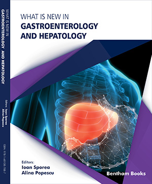
Gastroenterology and hepatology represent dynamic fields of study and practice in internal medicine, with numerous innovations manifesting over the last 30 years. What is New in Gastroenterology and Hepatology is a reference which presents updates on the latest advances in the aforementioned subspecialties. The book offers a broad range of topics in a structured, clear and comprehensive fashion. Thirty-three chapters provide knowledge on basic medical research and practical applications in the clinic with information contributed by several members of the Romanian Society of Gastroenterology and Hepatology (a medical society with a history of more than 60 years). The contents feature updates on common and rare disorders of the gastrointestinal tract (esophagus, stomach, intestines), pancreas and the liver, as well as new imaging techniques such as video capsule endoscopy and contrast enhanced ultrasound (CEUS). Special topics of recent interest such as the value of gut microbiota in medical interventions and the use of 'omics' technologies for precision medicine, and telemedicine are also included. What is New in Gastroenterology and Hepatology is an informative reference for all medical researchers and healthcare professionals (gastroenterologists, hepatologists, internal medicine physicians, surgeons, oncologists) who wish to keep themselves up to speed on new advances in these medical subspecialties.


