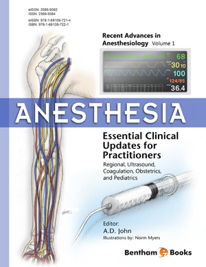
Book Volume 1
List of Contributors
Page: ii-iii (2)
Author: Atta- ur-Rahman and Shazia Anjum
DOI: 10.2174/9781608059942115010002
Safety Assessment of Mesenchymal Stem Cells in Musculoskeletal Implantation
Page: 3-29 (27)
Author: Greg Asatrian, Angel Pan, Michelle A. Scott and Aaron W. James
DOI: 10.2174/9781608059942115010003
PDF Price: $30
Abstract
There has been a rapid increase in our understanding in the isolation, culture and application of mesenchymal stem cells (MSC). Despite an increased understanding of MSC biology and new avenues for in vivo application - the standardization of laboratory practices for MSC is lacking. One particular example is in the examination of safety issues in MSC in vivo implantation. The following review will explore the current laboratory practices for the safety assessments of MSC implantation, from diverse viewpoints of such as histopathology, cytopathology, and cytogenetics. A snapshot of current practices and techniques is presented across three common types of MSC differentiation: bone, cartilage, and muscle tissue. Overall, we uncovered a relative lack of investigation of safety issues in MSC implantation. For example, cell proliferation and local inflammation were only assessed in less than one third of manuscripts. Additionally, the average length of study was less than two weeks, a short period limited in its detection of adverse outcomes. The present review uncovers a relative paucity of papers that place emphasis on safety outcomes for animal studies. Given the potential role of MSC in sarcomagenesis and other tumorigenesis, the routine performance safety assays for MSC mediated tissue engineering studies will facilitate the future translation to clinical use. Finally, we provide a set of practical preliminary suggestions is presented for safety assessments in MSC implantation models. In summary, in order to bridge the gap in translation from animal to human, increased practice and routinization of safety assessments in MSC implantation will be beneficial.
Strategies to Improve Immune Reconstitution After Haematopoietic Stem Cell Transplantation
Page: 30-64 (35)
Author: Guy Klamer, Sylvie Shen, Ning Xu, Tracy A. O`Brien and Alla Dolnikov
DOI: 10.2174/9781608059942115010004
PDF Price: $30
Abstract
Immunologic reconstitution is a critical component for successful outcome of haematopoietic stem cell transplantation. Chemotherapy and pre-transplant conditioning impairs thymic function leading to delayed T-cell regeneration and the increased risk of opportunistic infections and leukaemia relapse. Immune reconstitution can be promoted through administration of common γ-chain cytokines such as IL-2, IL-7 and IL-15. Prevention of thymic involution achieved by administration of keratinocyte growth factor, growth hormone and sex hormone inhibition has also been shown to improve immune reconstitution. Additionally, cell therapy that includes adoptive transfer of ex vivo generated T-cells or T-cell precursors, T-cells specific for viral or tumour antigens and, natural killer (NK) cells appears to be a promising therapeutic approach to improve immune reconstitution after transplantation. Pharmacological modulation of signalling pathways, such as Wnt and Notch, play an important role during different stages of Tcell development. Activation of Wnt signalling using small molecule inhibition of GSK3β was shown to promote post-transplant T-cell regeneration in pre-clinical models. The use of pharmaceutical agents to accelerate T-cell reconstitution and boost T-cell-mediated immunity in recipients of haematopoietic stem cell grafts warrants further investigation.
Alternative Biomaterial Substrates for Human Embryonic Stem Cell Culture
Page: 65-91 (27)
Author: Deepak Kumar, Ying Yang and Nicholas R. Forsyth
DOI: 10.2174/9781608059942115010005
PDF Price: $30
Abstract
The additive effect of stem cell therapy and biomaterial substrates provides exciting opportunities for tissue engineering and regenerative medicine applications. Nanofibrous substrates can be fabricated to mimic the nano-architectural structure of specific, native tissue extracellular matrix. This provides topographical structure and contact guidance, which can impact stem cell biology as well as direct their differentiation towards specific lineages. This chapter highlights nanofibrous substrates as an alternative tool for the expansion and differentiation of embryonic stem cells. Future applications of such technology could promote the use of hESC-derived cells for clinical applications.
Frontiers in Regenerative Medicine for Cornea and Ocular Surface
Page: 92-138 (47)
Author: Maria P. De Miguel, Ricardo P. Casaroli-Marano, Nuria Nieto-Nicolau, Eva M. Martínez-Conesa, Jorge L. Alió del Barrio, Jorge L. Alió, Sherezade Fuentes and Francisco Arnalich-Montiel
DOI: 10.2174/9781608059942115010006
PDF Price: $30
Abstract
The cornea represents two thirds of the eye’s refractive power and, together with the sclera, is the protective shield of intraocular structures. The cornea is composed of three cellular layers (epithelium, stroma and endothelium) and three acellular layers (Bowman’s, Dua’s and Descemet’s). Corneal pathologies can affect one or all corneal layers, producing corneal opacities. Penetrating keratoplasty is currently being displaced by lamellar techniques that selectively replace the diseased layer, but neither solves the classical difficulties encountered in corneal transplantation, such as immune rejection and a shortage of organs. The development of bioengineered corneas composed of prosthetic or natural scaffolds and autologous stem cells that differentiate into corneal cells could overcome these difficulties.
In recent years, much research has been carried out to find the optimal scaffold and the best source of stem cells to regenerate the corneal layers. Limbal stem cells (LSC) have arised as one of these sources, and the need to find a marker to distinguish them from more differentiated cells has also emerged. Both limbal and extraocular stem cells have been tested, and some techniques are already being used in clinical practice. These novel techniques for tissue engineering of functional corneal equivalents represent a new and fascinating way to treat corneal diseases. The new techniques allow us to treat patients with autologous grafts and can prevent the use of corneal stem cells, which are scarce and often unavailable.Subject Index
Page: 139-144 (6)
Author: Atta- ur-Rahman and Shazia Anjum
DOI: 10.2174/9781608059942115010007
Introduction
Stem cell and regenerative medicine research is a hot area of research which promises to change the face of medicine as it will be practiced in the years to come. Challenges in 21st century to combat cancer, Alzheimer and related diseases may well be addressed employing stem cell therapies and tissue regeneration. The first volume of ‘Frontiers in Stem Cell and Regenerative Medicine Research’ features reviews written by experts in key areas of stem cells and regenerative medicine. It summarizes the safety assessment of mesenchymal stem cells (MSC) in musculoskeletal implantation that can bridge the gap between translation from animals to humans. The most prevalent strategies to improve immune reconstitution after hematopoietic stem cell transplantation have also been focused upon. This is particularly important because chemotherapy and pre-transplant conditioning impairs thymic function. The application of regenerative medicine for repair of damaged cornea and ocular has also been discussed. The emerging techniques for tissue engineering of functional corneal equivalents represent a new and fascinating way to treat corneal diseases. The area of recently used nanofibrous substrates, as an alternative tool for the expansion and differentiation of embryonic stem cells, has been included in this e-book. In future, such technologies could promote the use of hESC-derived cells for clinical applications successfully.


